
-
Welcome to embodi3D Downloads! This is the largest and fastest growing library of 3D printable anatomic models generated from real medical scans on the Internet. A unique scientific resource, most of the material is free. Registered members can download, upload, and sell models. To convert your own medical scans to a 3D model, take a look at democratiz3D, our free and automated conversion service.
Alert (6/17/22) - The democratiz3D scan-to-model conversion app is down due to a technical issue. We are working on a solution.
Extremity, Lower (Leg)
Lower extremity: thigh, leg, ankle, foot.
1,289 files
-

Free
Right ankle, a 3D printable medical file of its normal anatomy converted from a CT scan DICOM dataset of a 75-year old female
By embodi3d
The ankle joint is a hinged synovial joint with primarily up-and-down movement (plantarflexion and dorsiflexion). However, when the range of motion of the ankle and subtalar joints (talocalcaneal and talocalcaneonavicular) is taken together, the complex functions as a universal joint.
The bony architecture of the ankle consists of three bones: the tibia, the fibula, and the talus. The articular surface of the tibia is referred to as the plafond. The medial malleolus is a bony process extending distally off the medial tibia. The distal-most aspect of the fibula is called the lateral malleolus. Together, the malleoli, along with their supporting ligaments, stabilize the talus underneath the tibia.
The bony arch formed by the tibial plafond and the two malleoli is referred to as the ankle "mortise" (or talar mortise). The mortise is a rectangular socket. The ankle is composed of three joints: the talocrural joint (also called talotibial joint, tibiotalar joint, talar mortise, talar joint), the subtalar joint (also called talocalcaneal), and the Inferior tibiofibular joint. The joint surface of all bones in the ankle are covered with articular cartilage.
This a 3D printable medical file converted from a CT scan DICOM dataset of a 75-year old female.
18 downloads
- ankle
- normal anatomy
- (and 6 more)
(0 reviews)0 comments
Updated
-

Free
Right leg and foot, a 3D printable STL file of their normal anatomy converted from a CT scan DICOM data of a 75-year old female
By embodi3d
The bones of the leg and foot form part of the appendicular skeleton that supports the many muscles of the lower limbs. These muscles work together to produce movements such as standing, walking, running, and jumping. At the same time, the bones and joints of the leg and foot must be strong enough to support the body’s weight while remaining flexible enough for movement and balance.
The tibia and fibulaare the bones that support the leg. The larger tibia or shinebone is located medial to the fibula and bears most of the weight.
At the superior (proximal) end of the tibia, a pair of flattened condyles articulate with the rounded condyles at the distal end of the femur to form the knee joint joint.
The tibia and fibula articulate at two sites. At the knee, a superior (proximal) tibiofibular joint is formed by the lateral tibial condyle and head of the fibula. At the ankle, an inferior (distal) tibiofibular joint is formed by the lower fibula and a lateral concavity (notch) on the lower tibia.
The feet are flexible structures of bones, joints, muscles, and soft tissues that let us stand upright and perform activities like walking, running, and jumping. The feet are divided into three sections:
-The forefoot contains the five toes (phalanges) and the five longer bones (metatarsals).
-The midfoot is a pyramid-like collection of bones that form the arches of the feet. These include the three cuneiform bones, the cuboid bone, and the navicular bone.
-The hindfoot forms the heel and ankle. The talus bone supports the leg bones (tibia and fibula), forming the ankle. The calcaneus (heel bone) is the largest bone in the foot.
24 downloads
- 3d imaging
- 3d printable medical file
- (and 8 more)
(0 reviews)0 comments
Updated
-

Free
Right knee joint, A 3D printable medical file of its normal anatomy converted from a CT scan DICOM dataset.
By embodi3d
The knee is the largest joint and one of the most important joints in the body. It plays an essential role in movement related to carrying the body weight in horizontal (running and walking) and vertical (jumping) directions.
The knee joint joins the thigh with the leg and consists of two articulations: one between the femur and tibia (tibiofemoral joint), and one between the femur and patella (patellofemoral joint). The knee is a modified hinge joint, which permits flexion and extension as well as slight internal and external rotation. The knee joint is vulnerable to injury and to the development of osteoarthritis.
The knee is composed of three functional compartments: the patellofemoral articulation, consisting of the patella, or "kneecap", and the patellar groove on the front of the femur through which it slides; and the medial and lateral tibiofemoral articulations linking the femur, or thigh bone, with the tibia, the main bone of the lower leg. The joint is bathed in synovial fluid which is contained inside the synovial membrane called the joint capsule.
This is a 3D-printable medical STL file of normal right knee joint converted from a CT scan DICOM dataset of a 75-year old female patient(STS-016).
35 downloads
- normal anatomy
- knee
- (and 9 more)
(0 reviews)0 comments
Updated
-

Free
Left knee joint, A 3D printable medical file of its normal anatomy converted from a CT scan DICOM dataset.
By embodi3d
The knee is the largest joint and one of the most important joints in the body. It plays an essential role in movement related to carrying the body weight in horizontal (running and walking) and vertical (jumping) directions.
The knee joint joins the thigh with the leg and consists of two articulations: one between the femur and tibia (tibiofemoral joint), and one between the femur and patella (patellofemoral joint). The knee is a modified hinge joint, which permits flexion and extension as well as slight internal and external rotation. The knee joint is vulnerable to injury and to the development of osteoarthritis.
The knee is composed of three functional compartments: the patellofemoral articulation, consisting of the patella, or "kneecap", and the patellar groove on the front of the femur through which it slides; and the medial and lateral tibiofemoral articulations linking the femur, or thigh bone, with the tibia, the main bone of the lower leg. The joint is bathed in synovial fluid which is contained inside the synovial membrane called the joint capsule.
This is a 3D-printable medical STL file of normal left knee joint converted from a CT scan dataset of a 75-year old female patient (STS-016).
13 downloads
- normal anatomy
- knee
- (and 7 more)
(0 reviews)0 comments
Updated
-
Free
A 3D-printable medical file of a right thigh soft tissue sarcoma, converted from a ct scan DICOM dataset
By embodi3d
This is a 3D model of a right thigh soft tissue mass extracted from the ct scan DICOM dataset of a 62-year old female. Histopathological examination revealed the mass to be sclerosing epithelioid fibrosarcoma of intermediate grade. 14 days prior to the pathological examination, MRI was done for this patient and 40 days after the diagnosis a PET scan was done.Her treatment plan was a combination of surgical resection of the mass and radiotherapy. Bone metastasis was confirmed 235 days after the initial diagnosis.The patient died 428 days after the diagnosis of the bone metastasis. (STS-012)
0 downloads
- sarcoma
- soft tissue
- (and 6 more)
(0 reviews)0 comments
Updated
-

Free
The femoral head and neck , a 3D-printable medical file of their normal anatomy converted from a CT scan DICOM dataset
By embodi3d
The uppermost part of the femur consists of the head which is the highest part, and the neck which is a flattened pyramidal process that connects the head with the shaft forming a wide angle with the later. The superior border of the neck is short and thick and ends with the greater trochanter, while the inferior border is longer and ends with the lesser trochanter. The blood supply enters through the neck. This is a 3D-printable medical STL file converted from a CT scan DICOM dataset.
3 downloads
- femur
- femoral head
- (and 4 more)
(0 reviews)0 comments
Updated
-

Free
lower extremity Left Leg Bone Model 3D Printable STL File Converted from CT Scan
By embodi3d
This is the normal left leg bone model (including foot) of a 56 year old male with right anterior thigh pleomorphic leiomyosarcoma. This is an STL file created from DICOM images of his CT scan which may be used for 3D printing.
The leg includes the area between the knee and the ankle and houses the tibia and fibula. The proximal tibia includes the medial plateau (which is concave) and the lateral plateau (which is convex). The Proximal tibia has a 7-10 degree posterior slope. The tibial tuberosity is located on the anterior proximal tibia, which is where the patellar tendon attaches. On the anteromedial surface of the tibia is Gerdy's tubercle, where the sartorius, gracilis, and semitendinosus attach. The distal tibia creates the superior and medial (plafond and medial malleolus) of the ankle joint.
The proximal fibula is the attachment for the posterolateral corner structures of the knee joint. The peroneal nerve wraps around the fibular neck. The distal fibula is the lateral malleolus and a common site for ankle fractures.
This model was created from the file STS_014.
8 downloads
(0 reviews)0 comments
Updated
-

Free
leg Right Leg Bone Model 3D Printable STL File Converted from CT Scan
By embodi3d
This is the normal right leg bone model (including foot) of a 56 year old male with right anterior thigh pleomorphic leiomyosarcoma. This is an STL file created from DICOM images of his CT scan which may be used for 3D printing.
The leg includes the area between the knee and the ankle and houses the tibia and fibula. The proximal tibia includes the medial plateau (which is concave) and the lateral plateau (which is convex). The Proximal tibia has a 7-10 degree posterior slope. The tibial tuberosity is located on the anterior proximal tibia, which is where the patellar tendon attaches. On the anteromedial surface of the tibia is Gerdy's tubercle, where the sartorius, gracilis, and semitendinosus attach. The distal tibia creates the superior and medial (plafond and medial malleolus) of the ankle joint.
The proximal fibula is the attachment for the posterolateral corner structures of the knee joint. The peroneal nerve wraps around the fibular neck. The distal fibula is the lateral malleolus and a common site for ankle fractures.
This model was created from the file STS_014.
5 downloads
- bone
- bone model
- (and 10 more)
(0 reviews)0 comments
Updated
-

Free
knee Left Knee Bone Model 3D Printable STL File Converted from CT Scan
By embodi3d
This is the normal left knee bone model of a 56 year old male with right anterior thigh pleomorphic leiomyosarcoma. This is an STL file created from DICOM images of his CT scan which may be used for 3D printing.
The knee is composed of 3 separate joints: two hinge joints (medial and lateral femorotibial joints), and one sellar, or gliding, joint (the patellofemoral joint). These also compose the three compartments of the knee: medial, lateral, and patellofemoral. Although the knee is thought of as a hinge joint, it actually has 6 degrees of motion: extension/flexion, internal/external rotation, varus/valgus, anterior/posterior translation, medial/lateral translation, and compression/distraction. In order to provide stability to this inherently unstable knee, static and dynamic stabilizers surround the knee, including muscles and ligaments.
The proximal tibia includes the medial plateau (which is concave) and the lateral plateau (which is convex). The Proximal tibia has a 7-10 degree posterior slope. The tibial tuberosity is located on the anterior proximal tibia, which is where the patellar tendon attaches. On the anteromedial surface of the tibia is Gerdy's tubercle, where the sartorius, gracilis, and semitendinosus attach. The proximal fibula is the attachment for the posterolateral corner structures of the knee joint. The peroneal nerve wraps around the fibular neck.
This model was created from the file STS_014.
49 downloads
- bone
- bone model
- (and 10 more)
(0 reviews)0 comments
Updated
-

Free
foot and ankle Normal Right Foot and Ankle Bone Model 3D Printable STL File Converted from CT Scan
By embodi3d
This is the normal right foot and ankle bone model of a 56 year old male with right anterior thigh pleomorphic leiomyosarcoma. This is an STL file created from DICOM images of his CT scan which may be used for 3D printing.
The ankle is a hinge (or ginglymus) joint made of the distal tibia (tibial plafond, medial and posterior malleoli) superiorly and medially, the distal fibula (lateral malleolus) laterally and the talus inferiorly. Together, these structures form the ankle “mortise”, which refers to the bony arch. The normal range of motion is 20 degrees dorsiflexion and 50 degrees plantarflexion. Stability is provided by the anterior talofibular ligament (ATFL), calcaneofibular ligament (CFL), and posterior talofibular ligament (PTFL) laterally, and the superficial and deep deltoid ligaments medially. The ankle is one of my most common sites of musculoskeletal injury, including ankle fractures and ankle sprains, due to the ability of the joint to invert and evert. The most common ligament involved in the ATFL.
The foot is commonly divided into three segments: hindfoot, midfoot, and forefoot. These sections are divided by the transverse tarsal joint (between the talus and calcaneus proximally and navicular and cuboid distally), and the tarsometatarsal joint (between the cuboids and cuneiforms proximally and the metatarsals distally). The first tarsometatarsal joint (medially) is termed the “Lisfranc” joint and is the site of the Lisfranc injury seen primarily in athletic injuries.
This model was created from the file STS_014.
12 downloads
(0 reviews)0 comments
Updated
-
foot and ankle Normal Left Foot and Ankle Bone Model 3D Printable STL File Converted from CT Scan
By embodi3d
This is the normal right foot and ankle bone model of a 56 year old male with right anterior thigh pleomorphic leiomyosarcoma. This is an STL file created from DICOM images of his CT scan which may be used for 3D printing.
The ankle is a hinge (or ginglymus) joint made of the distal tibia (tibial plafond, medial and posterior malleoli) superiorly and medially, the distal fibula (lateral malleolus) laterally and the talus inferiorly. Together, these structures form the ankle “mortise”, which refers to the bony arch. The normal range of motion is 20 degrees dorsiflexion and 50 degrees plantarflexion. Stability is provided by the anterior talofibular ligament (ATFL), calcaneofibular ligament (CFL), and posterior talofibular ligament (PTFL) laterally, and the superficial and deep deltoid ligaments medially. The ankle is one of my most common sites of musculoskeletal injury, including ankle fractures and ankle sprains, due to the ability of the joint to invert and evert. The most common ligament involved in the ATFL.
The foot is commonly divided into three segments: hindfoot, midfoot, and forefoot. These sections are divided by the transverse tarsal joint (between the talus and calcaneus proximally and navicular and cuboid distally), and the tarsometatarsal joint (between the cuboids and cuneiforms proximally and the metatarsals distally). The first tarsometatarsal joint (medially) is termed the “Lisfranc” joint and is the site of the Lisfranc injury seen primarily in athletic injuries.
This model was created from the file STS_014.
28 downloads
(0 reviews)0 comments
Updated
-

Free
leg Right Leg Bone Model 3D Printable STL File Converted from CT Scan
By embodi3d
This is the normal right leg bone model (including foot) of an 82-year-old male. This is an STL file created from DICOM images of his CT scan which may be used for 3D printing.
The leg includes the area between the knee and the ankle and houses the tibia and fibula. The proximal tibia includes the medial plateau (which is concave) and the lateral plateau (which is convex). The Proximal tibia has a 7-10 degree posterior slope. On the anterior proximal tibia, the tibial tuberosity, where the patellar tendon attaches. On the anteromedial surface of the tibia is Gerdy's tubercle, where the sartorius, gracilis, and semitendinosus attach. The distal tibia creates the superior and medial (plafond and medial malleolus) of the ankle joint.
The proximal fibula is the attachment for the posterolateral corner structures of the knee joint. The peroneal nerve wraps around the fibular neck. The distal fibula is the lateral malleolus and a common site for ankle fractures.
This model was created from the file STS_013.
16 downloads
(0 reviews)0 comments
Updated
-

Free
hip Normal Right Hip Bone Model from STL File from CT Scan for 3D Printing
By embodi3d
This is the normal right hip model of an 82-year-old male. This is an STL file created from DICOM images of his CT scan which may be used for 3D printing.
The hip joint is a ball and socket joint that has intrinsic stability from osseous, ligamentous, and muscular structures. The hip capsule is made of the iliofemoral, pubofemoral, and ischiofemoral ligaments which attach from the acetabulum to the femoral neck. The normal acetabulum is anteverted 15 degrees and abducted 45 degrees. The normal femoral anteversion is between 10-15 degrees. The proximal femur also includes the greater trochanter, to which the external rotators are attached, and the lesser trochanter, to which the iliopsoas is attached.
This model was created from the file STS_013.
14 downloads
- normal anatomy
- 3d printable
- (and 6 more)
(0 reviews)0 comments
Updated
-

Free
dedifferentiated liposarcoma Left Thigh Dedifferentiated Liposarcoma 3D Printable Bone Model STL File Converted from CT Scan
By embodi3d
This image is the left thigh bone rendering of an 82 year old male with a dedifferentiated liposarcoma in the anterior compartment of the left thigh. This is an STL file created from DICOM images of his CT scan which may be used for 3D printing.
Liposarcomas are the second most common soft tissue sarcoma in adults, occurring more commonly in males between the ages of 50 and 80 years old. They present as a slow growing, painless mass typically located in the extremities, with the thigh being the most common location. Multiple variants of liposarcomas exist, but the dedifferentiated type is a high-grade sarcoma. Dedifferentiated liposarcomas are typically located adjacent to a well-differentiated lipomatous lesion. The incidence of pulmonary metastasis increases with grade. Therefore, work up of the lesion consists of MRI, biopsy through the area of future resection, CT of the chest, abdomen, and pelvis to rule out metastases. Treatment consists of radiation and wide surgical resection but chemotherapy agents are being developed to target chromosomal abnormalities associated with certain well-differentiated and dedifferentiated liposarcomas. This patient received radiation and resection of the tumor and has not had a metastasis or recurrence in 4.5 years.
This image came from the file STS_013.
21 downloads
- 3d medical printing
- 3d printable
- (and 14 more)
(0 reviews)0 comments
Updated
-

Free
(0 reviews)0 comments
Submitted
-

Free
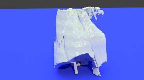
Lower body - processed
By shunt1
Lower body - processed, femoral, common, artery, superficial, stl, pelvis, hip, lower, limb, knee, patella, tibia, fibula, 3d, model, bone
12 downloads
(0 reviews)0 comments
Updated
-

Free
(0 reviews)0 comments
Updated
-

Free
Displasia cadera con rodilla - processed
By Victor Barro
Displasia cadera con rodilla - processed
0 downloads
(0 reviews)0 comments
Updated
-

Free
Instructables 3D model of the leg bones - processed
By mitchell
Instructables 3D model of the leg bones - processed
1 download
(0 reviews)0 comments
Updated
-

Free
jamsheed
By jamsheed
i want only knee joint in stl format and it should be processed for rpt machine
STS_010-right-knee_nrrd.a4c03b8b73f8bfa362123e673bd23d7f_IYNADG.stl
femur, knee, patella, lower, limb, stl, 3d, model, tibia, fibula, bone, printable
6 downloads
(0 reviews)0 comments
Updated
-

Free
knee joint stl
By jamsheed
i want to make 3d modelling of knee joint
STS_010-right-knee_nrrd.a4c03b8b73f8bfa362123e673bd23d7f_IYNADG.stl
femur, knee, patella, lower, limb, stl, 3d, model, tibia, fibula, bone, printable
12 downloads
(0 reviews)0 comments
Updated
-

Free
(0 reviews)0 comments
Submitted
-
(0 reviews)
0 comments
Submitted
-

Free
(0 reviews)0 comments
Submitted
-

Free
knee - processed
By willyci
knee - processed, lower, limb, stl, knee, joint, skin, fémur, tibia, fibula, 3d, model, printable
0 downloads
(0 reviews)0 comments
Updated


-
File Reviews
-
File Comments
-
Recent Forum Posts
-

By jangiddrrk · Posted
Free connections, find your partner with no obligations Verified Damsels [URL=https://datesnow.life]Outstanding Сasual Dating[/URL] -
 Hello everyone, I hope this message finds you well. I am reaching out to our community in hopes of finding assistance with a project I'm currently undertaking. Specifically, I am in need of a detailed 3D model of the temporomandibular joint (TMJ). If anyone in our community has access to or expertise in creating high-quality 3D models, particularly of the temporomandibular joint, I would greatly appreciate any assistance you can offer. Whether you have a model readily available or can provide guidance on where to find one, your contribution would be invaluable to my project. Thank you in advance for your assistance and support.
 -

-

-

-


