
-
Welcome to embodi3D Downloads! This is the largest and fastest growing library of 3D printable anatomic models generated from real medical scans on the Internet. A unique scientific resource, most of the material is free. Registered members can download, upload, and sell models. To convert your own medical scans to a 3D model, take a look at democratiz3D, our free and automated conversion service.
Alert (6/17/22) - The democratiz3D scan-to-model conversion app is down due to a technical issue. We are working on a solution.
Extremity, Lower (Leg)
Lower extremity: thigh, leg, ankle, foot.
1,289 files
-

Free
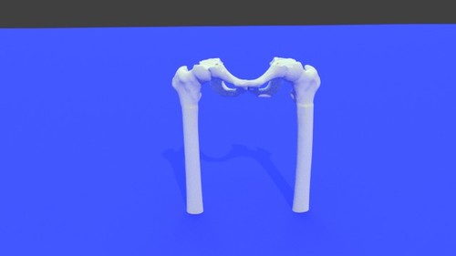
Left Hip Joint 3D Printable STL File Converted From CT Scan
By embodi3d
The hip joint is a large synovial socket and ball joint which is formed by the femoral head (the ball) and the acetabulum (the socket). The acetabulum is formed by pelvic bones; the ilium, the ischium and the pubis. The hip joint represents the articulation between the lower extremity and the axial skeleton and allows a high degree of mobility while being stable. This 3D model was created from the file STS_036 The original CT examination can be reviewed at: The 3D muscle model created from this scan can be reviewed at:2 downloads
(0 reviews)0 comments
Updated
-
Free
(0 reviews)0 comments
Updated
-

Free
Moose1 - Radius & Ulna - stl file processed
By Mission 3D
Moose1 - Radius & Ulna - stl file processed1 download
(0 reviews)0 comments
Submitted
-

Free
Moose1 - Radius & Ulna - Detailed STL - stl file processed
By Mission 3D
Moose1 - Radius & Ulna - Detailed STL - stl file processed0 downloads
(0 reviews)0 comments
Submitted
-

Free
Moose - Radius & Ulna - stl file processed
By Mission 3D
Moose - Radius & Ulna - stl file processed1 download
(0 reviews)0 comments
Submitted
-

Free
(0 reviews)0 comments
Updated
-

Free
Left Knee Joint 3D Printable STL File Converted From CT Scan
By embodi3d
Left Knee Joint 3D Printable STL File Converted From CT Scan - stl file processed
The knee joint is formed by three bones: the femur, the tibia and the patella. the knee joint is the largest synovial joint and provides the flexion and extension movements of the leg as well as relative medial and lateral rotations while in relative flexion.
The knee joint articulations are two condylar joints between the femur and the tibia as well as a joint between the patella and the femur. Although the fibula is closely related to the knee joint but it doesn't share in articulation. The knee joint is also formed by some ligaments and cartilage called (menisci) which are best imaged by MRI. This 3D model was created from the file STS_045. The source CT scan used to create this model can be found here.186 downloads
- knee
- knee joint
- (and 11 more)
(1 review)0 comments
Updated
-

Free
Left Hip Joint 3D Printable STL File Converted From CT Scan
By embodi3d
The hip joint is a large synovial socket and ball joint which is formed by the femoral head (the ball) and the acetabulum (the socket). The acetabulum is is formed by pelvic bones; the ilium, the ischium and the pubis. The hip joint represents the articulation between the lower extremity and the axial skeleton and allows a high degree of mobility while being stable. This 3D model was created from the file STS_044. The source CT scan used to create this model can be found here.27 downloads
(0 reviews)0 comments
Updated
-

Free
Right Hip Joint 3D Printable STL File Converted From CT Scan
By embodi3d
The hip joint is a large synovial socket and ball joint which is formed by the femoral head (the ball) and the acetabulum (the socket). The acetabulum is is formed by pelvic bones; the ilium, the ischium and the pubis. The hip joint represents the articulation between the lower extremity and the axial skeleton and allows a high degree of mobility while being stable. This 3D model was created from the file STS_044. The source CT scan used to create this model can be found here.74 downloads
(0 reviews)0 comments
Updated
-

Free
Right Knee Joint 3D Printable STL File Converted From CT Scan
By embodi3d
The knee joint is formed by three bones: the femur, the tibia and the patella. the knee joint is the largest synovial joint and provides the flexion and extension movements of the leg as well as relative medial and lateral rotations while in relative flexion.
The knee joint articulations are two condylar joints between the femur and the tibia as well as a joint between the patella and the femur. Although the fibula is closely related to the knee joint but it doesn't share in articulation. The knee joint is also formed by some ligaments and cartilage called (menisci) which are best imaged by MRI. This 3D model was created from the file STS_045. The source scan be be found here.95 downloads
- knee
- knee joint
- (and 8 more)
(0 reviews)0 comments
Updated
-

Free
Left Adductor Muscle Extraskeletal Osteosarcoma 3D Printable STL File Converted From CT Scan
By embodi3d
This 3D model represents a case of high grade extraskeletal osteosarcoma affecting the left adductor muscle of a 27 years old male. The patient was treated by surgical excision follower by chemotherapy. A cross sectional CT image is attached showing the lesion in axial, coronal and sagittal planes. Extraskeletal osteosarcoma (ESOS) is one of the rare malignant neoplasms that affects the mesenchymal tissues such as the retroperitoneum as well as the soft tissue of the extremities with no significant connection to the related bones. Extraskeletal osteosarcoma usually affects people between 40 years and 80 years and is more common in males with a documented risk factor which is radiation exposure. The common presentation is enlarged or swollen soft tissue which could be painful or not. Extraskeletal osteosarcoma is diagnosed by plain x-ray, CT or MRI as the soft tissue shows variable calcification. The most common affected sites are the lower extremities followed by upper extremities and retroperitoneum. Most of patients are presented with metastasis at time of diagnosis which leads to a generally poor prognosis. The usual treatment is surgical excision of the primary tumor as the tumor is insensitive to chemotherapy or radiotherapy. This 3D model was created from the file STS_045. The source scan can be found here.11 downloads
- osteosarcoma
- extraskeletal osteosarcoma
- (and 11 more)
(0 reviews)0 comments
Updated
-

Free
(0 reviews)0 comments
Submitted
-

Free
(0 reviews)0 comments
Updated
-

Free
(0 reviews)0 comments
Updated
-

Free
lesser leg - stl file processed
By sstaal47
lesser leg - stl file processed
0 downloads
- lesser leg
- 3dmodel
- (and 5 more)
(0 reviews)0 comments
Updated
-

Free
BC, remaining bone, no femur - stl file processed
By Felasfa
BC, remaining bone, no femur - stl file processed
14 downloads
(0 reviews)0 comments
Updated
-

Free
resected bone, including femur - stl file processed
By Felasfa
resected bone, including femur - stl file processed1 download
(0 reviews)0 comments
Submitted
-

Free
Muestra - stl file processed
Muestra - stl file processed
pelvis, stl, lower limb, bones, 3dmodel
5 downloads
- pelvis
- lower limb
- (and 3 more)
(0 reviews)0 comments
Updated
-

Free
8 Bone 2.0 Sagittal.nrrd - stl file processed
By Rfperez
8 Bone 2.0 Sagittal.nrrd - stl file processed
3 downloads
(0 reviews)0 comments
Updated
-

Free
8 Bone 2.0 Sagittal.nrrd - stl file processed
By Rfperez
8 Bone 2.0 Sagittal.nrrd - stl file processed, foot, ankle, tibia, fibula, scaphoid, cuboid, tarsal, metatarsal, lower, limb, joint, bone, printable, 3d, model
10 downloads
(0 reviews)0 comments
Updated
-
(0 reviews)
0 comments
Submitted
-

Free
(0 reviews)0 comments
Submitted
-

Free
(0 reviews)0 comments
Submitted
-

Free
(0 reviews)0 comments
Submitted
-

Free
left femur top half - stl file processed
By bryanc
left femur top half - stl file processed
9 downloads
- femur
- lower limb
- (and 3 more)
(0 reviews)0 comments
Updated


-
File Reviews
-
File Comments
-
Recent Forum Posts
-

By jangiddrrk · Posted
Free connections, find your partner with no obligations Verified Damsels [URL=https://datesnow.life]Outstanding Сasual Dating[/URL] -
 Hello everyone, I hope this message finds you well. I am reaching out to our community in hopes of finding assistance with a project I'm currently undertaking. Specifically, I am in need of a detailed 3D model of the temporomandibular joint (TMJ). If anyone in our community has access to or expertise in creating high-quality 3D models, particularly of the temporomandibular joint, I would greatly appreciate any assistance you can offer. Whether you have a model readily available or can provide guidance on where to find one, your contribution would be invaluable to my project. Thank you in advance for your assistance and support.
 -

-

-

-


