
-
Welcome to embodi3D Downloads! This is the largest and fastest growing library of 3D printable anatomic models generated from real medical scans on the Internet. A unique scientific resource, most of the material is free. Registered members can download, upload, and sell models. To convert your own medical scans to a 3D model, take a look at democratiz3D, our free and automated conversion service.
Alert (6/17/22) - The democratiz3D scan-to-model conversion app is down due to a technical issue. We are working on a solution.
Lower Extremity CTs
CTs of the hip, thigh, leg, and foot
712 files
-

Free
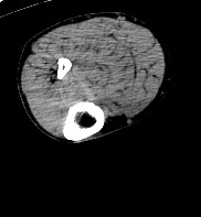
mi rodilla izq
By tito matus
nrrd to stl
ct without contrast, knee, lower limb, stl, axial, ct
1 download
- ct without contrast
- knee
- (and 4 more)
(0 reviews)0 comments
Updated
-

Free
Lower Limbs - CT scan
The lower limb consists of the hip joint, thigh, knee joint, leg, ankle joint and the foot. The thigh is the upper segment of the lower limb extending from the pelvis down to the knee joint. It contains the longest bone in the body which is the femur with multiple muscles,vascular structure and nerves. The knee joint is formed by three bones: the femur, the tibia and the patella. the knee joint is the largest synovial joint and provides the flexion and extension movements of the leg as well as relative medial and lateral rotations while in relative flexion.
The knee joint articulations are two condylar joints between the femur and the tibia as well as a joint between the patella and the femur. Although the fibula is closely related to the knee joint but it doesn't share in articulation. The leg is the lower limb segment extends from the knee joint down to the ankle joint. It is composed of two bones: the tibia and the fibula with numerous muscles and ligaments. The ankle joint is comprised of the tibia, fibula, talus, and calcaneus as well as the supporting ligaments, muscles and neurovascular bundles. The ankle is a synovial joint composed of the distal tibia and fibula as they articulate with the talus. The distal tibia and fibula articulate with each other at the distal tibiofibular joint which is more commonly referred to as the tibiofibular syndesmosis. The foot is a highly developed, biomechanically complex structure that serves to bear the weight of the body. The foot can be divided into 3 parts: the hindfoot, the midfoot, and the forefoot. The hindfoot is composed of 2 of the 7 tarsal bones, the talus, and the calcaneus; the midfoot contains the rest of the tarsal bones; and the forefoot contains the metatarsals and the phalanges. The CT scan is derived from the file STS_041 The 3D bone model created from this scan can be reviewed at: The 3D muscle model created from this scan can be reviewed at: The 3D skin model created from this scan can be reviewed at:3 downloads
(0 reviews)0 comments
Updated
-

Free
Left knee - CT scan
By embodi3d
The knee joint is formed by three bones: the femur, the tibia and the patella. the knee joint is the largest synovial joint and provides the flexion and extension movements of the leg as well as relative medial and lateral rotations while in relative flexion.
The knee joint articulations are two condylar joints between the femur and the tibia as well as a joint between the patella and the femur. Although the fibula is closely related to the knee joint but it doesn't share in articulation. The knee joint is also formed by some ligaments and cartilage called (menisci) which are best imaged by MRI. The CT scan is derived from the file STS_051 The 3D bone model created from this scan can be reviewed at: The 3D muscle model created from this scan can be reviewed at: The 3D skin model created from this scan can be reviewed at:51 downloads
(0 reviews)0 comments
Updated
-

Free
Right knee - CT scan
By embodi3d
The knee joint is formed by three bones: the femur, the tibia and the patella. the knee joint is the largest synovial joint and provides the flexion and extension movements of the leg as well as relative medial and lateral rotations while in relative flexion.
The knee joint articulations are two condylar joints between the femur and the tibia as well as a joint between the patella and the femur. Although the fibula is closely related to the knee joint but it doesn't share in articulation. The knee joint is also formed by some ligaments and cartilage called (menisci) which are best imaged by MRI. The CT scan is derived from the file STS_051 The 3D bone model created from this scan can be reviewed at: The 3D muscle model created from this scan can be reviewed at: The 3D skin model created from this scan can be reviewed at:79 downloads
(0 reviews)0 comments
Updated
-

Free
Left Ankle - CT scan
By embodi3d
The ankle joint is comprised of the tibia, fibula, talus, and calcaneus as well as the supporting ligaments, muscles and neurovascular bundles.
The ankle is a synovial joint composed of the distal tibia and fibula as they articulate with the talus. The distal tibia and fibula articulate with each other at the distal tibiofibular joint which is more commonly referred to as the tibiofibular syndesmosis. The CT scan is derived from the file STS_051 The 3D bone model created from this scan can be reviewed at: The 3D muscle model created from this scan can be reviewed at: The 3D skin model created from this scan can be reviewed at:14 downloads
(0 reviews)0 comments
Updated
-

Free
Right Ankle - CT scan
By embodi3d
The ankle joint is comprised of the tibia, fibula, talus, and calcaneus as well as the supporting ligaments, muscles and neurovascular bundles. The ankle is a synovial joint composed of the distal tibia and fibula as they articulate with the talus. The distal tibia and fibula articulate with each other at the distal tibiofibular joint which is more commonly referred to as the tibiofibular syndesmosis. The CT scan is derived from the file STS_051 The 3D bone model created from this scan can be reviewed at: The 3D muscle model created from this scan can be reviewed at: The 3D skin model created from this scan can be reviewed at:15 downloads
(0 reviews)0 comments
Updated
-

Free
(0 reviews)0 comments
Updated
-
Free
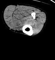
Left knee - CT scan
By embodi3d
The knee joint is formed by three bones: the femur, the tibia and the patella. the knee joint is the largest synovial joint and provides the flexion and extension movements of the leg as well as relative medial and lateral rotations while in relative flexion.
The knee joint articulations are two condylar joints between the femur and the tibia as well as a joint between the patella and the femur. Although the fibula is closely related to the knee joint but it doesn't share in articulation. The knee joint is also formed by some ligaments and cartilage called (mensci) which are best imaged by MRI. The CT scan is derived from the file STS_039 The 3D bone model created from this scan can be reviewed at: The 3D muscle model created from this scan can be reviewed at: The 3D skin model created from this scan can be reviewed at:51 downloads
- knee joint
- femur
- (and 5 more)
(0 reviews)0 comments
Updated
-
Free
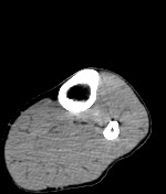
Right knee - CT scan
By embodi3d
The knee joint is formed by three bones: the femur, the tibia and the patella. the knee joint is the largest synovial joint and provides the flexion and extension movements of the leg as well as relative medial and lateral rotations while in relative flexion.
The knee joint articulations are two condylar joints between the femur and the tibia as well as a joint between the patella and the femur. Although the fibula is closely related to the knee joint but it doesn't share in articulation. The knee joint is also formed by some ligaments and cartilage called (mensci) which are best imaged by MRI. The CT scan is derived from the file STS_039 The 3D bone model created from this scan can be reviewed at: The 3D muscle model created from this scan can be reviewed at: The 3D skin model created from this scan can be reviewed at:278 downloads
- knee joint
- femur
- (and 5 more)
(0 reviews)0 comments
Updated
-

Free
Left foot - CT scan
By embodi3d
The foot is a highly developed, biomechanically complex structure that serves to bear the weight of the body.
The foot can be divided into 3 parts: the hindfoot, the midfoot, and the forefoot. The hindfoot is composed of 2 of the 7 tarsal bones, the talus, and the calcaneus; the midfoot contains the rest of the tarsal bones; and the forefoot contains the metatarsals and the phalanges. The CT scan is derived from the file STS_039 The 3D bone model created from this scan can be reviewed at: The 3D muscle model created from this scan can be reviewed at: The 3D skin model created from this scan can be reviewed at:20 downloads
(0 reviews)0 comments
Updated
-

Free
Right foot - CT scan
By embodi3d
The foot is a highly developed, biomechanically complex structure that serves to bear the weight of the body. The foot can be divided into 3 parts: the hindfoot, the midfoot, and the forefoot. The hindfoot is composed of 2 of the 7 tarsal bones, the talus, and the calcaneus; the midfoot contains the rest of the tarsal bones; and the forefoot contains the metatarsals and the phalanges. The CT scan is derived from the file STS_039 The 3D bone model created from this scan can be reviewed at: The 3D muscle model created from this scan can be reviewed at: The 3D skin model created from this scan can be reviewed at:13 downloads
(0 reviews)0 comments
Updated
-

Free
Left Hip - CT scan
By embodi3d
The hip joint is a large synovial socket and ball joint which is formed by the femoral head (the ball) and the acetabulum (the socket). The acetabulum is formed by pelvic bones; the ilium, the ischium and the pubis. The hip joint represents the articulation between the lower extremity and the axial skeleton and allows a high degree of mobility while being stable. The CT scan is derived from the file STS_040 The 3D bone model created from this scan can be reviewed at: The 3D muscle model created from this scan can be reviewed at:5 downloads
- hip joint
- socket and ball joint
- (and 8 more)
(0 reviews)0 comments
Updated
-
Free
Right Hip - CT scan
By embodi3d
The hip joint is a large synovial socket and ball joint which is formed by the femoral head (the ball) and the acetabulum (the socket). The acetabulum is formed by pelvic bones; the ilium, the ischium and the pubis. The hip joint represents the articulation between the lower extremity and the axial skeleton and allows a high degree of mobility while being stable. The CT scan is derived from the file STS_040 The 3D bone model created from this scan can be reviewed at: The 3D muscle model created from this scan can be reviewed at:5 downloads
- hip joint
- socket and ball joint
- (and 8 more)
(0 reviews)0 comments
Updated
-

Free
(0 reviews)0 comments
Updated
-

Free
Left Hip - CT scan
By embodi3d
The hip joint is a large synovial socket and ball joint which is formed by the femoral head (the ball) and the acetabulum (the socket). The acetabulum is formed by pelvic bones; the ilium, the ischium and the pubis. The hip joint represents the articulation between the lower extremity and the axial skeleton and allows a high degree of mobility while being stable. The CT scan is derived from the file STS_037 The 3D bone model created from this scan can be reviewed at: The 3D muscle model created from this scan can be reviewed at:3 downloads
(0 reviews)0 comments
Updated
-

Free
Right Hip - CT scan
By embodi3d
The hip joint is a large synovial socket and ball joint which is formed by the femoral head (the ball) and the acetabulum (the socket). The acetabulum is formed by pelvic bones; the ilium, the ischium and the pubis. The hip joint represents the articulation between the lower extremity and the axial skeleton and allows a high degree of mobility while being stable. The CT scan is derived from the file STS_037 The 3D bone model created from this scan can be reviewed at: The 3D muscle model created from this scan can be reviewed at:5 downloads
(0 reviews)0 comments
Updated
-

Free
Left Hip Joint CT scan
By embodi3d
The hip joint is a large synovial socket and ball joint which is formed by the femoral head (the ball) and the acetabulum (the socket). The acetabulum is formed by pelvic bones; the ilium, the ischium and the pubis. The hip joint represents the articulation between the lower extremity and the axial skeleton and allows a high degree of mobility while being stable. This CT scan was cropped from the file STS_036 The 3D bone model created from this scan can be reviewed at: The 3D muscle model created from this scan can be reviewed at:1 download
- hip
- socket and ball joint
- (and 9 more)
(0 reviews)0 comments
Updated
-

Free
Right Hip CT scan
By embodi3d
The hip joint is a large synovial socket and ball joint which is formed by the femoral head (the ball) and the acetabulum (the socket). The acetabulum is formed by pelvic bones; the ilium, the ischium and the pubis.
The hip joint represents the articulation between the lower extremity and the axial skeleton and allows a high degree of mobility while being stable. This CT scan was cropped from the file STS_036 The 3D bone model created from this scan can be reviewed at: The 3D muscle model created from this scan can be reviewed at:0 downloads
(0 reviews)0 comments
Updated
-

Free
(0 reviews)0 comments
Updated
-

Free
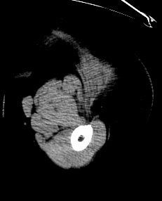
Left Groin Leiomyosarcoma CT Scan
By embodi3d
Left Groin Leiomyosarcoma 3D Printable STL File Converted From CT Scan This 3D model was created from the file STS_036
A 3D printable STL file model created from this scan can be found here.
4 downloads
(0 reviews)0 comments
Updated
-

Free
(0 reviews)0 comments
Updated
-
Free
Left Knee Joint CT Scan
By embodi3d
Left Knee Joint 3D Printable STL File Converted From CT Scan
This 3D model was created from the file STS_045
A 3D printable STL file model created from this scan can be found here.
31 downloads
(0 reviews)0 comments
Updated
-

Free
Left Hip Joint 3D Printable STL File Converted From CT Scan
By embodi3d
Left Hip Joint 3D Printable STL File Converted From CT Scan
This 3D model was created from the file STS_044. A 3D printable STL file model created from this scan can be found here.
7 downloads
(0 reviews)0 comments
Updated
-

Free
Right Hip Joint 3D Printable STL File Converted From CT Scan
By embodi3d
Right Hip Joint 3D Printable STL File Converted From CT Scan
This 3D model was created from the file STS_044. A 3D printable STL file model created from this scan can be found here.
9 downloads
(0 reviews)0 comments
Updated
-

Free
Right Knee Joint 3D Printable STL File Converted From CT Scan
By embodi3d
This 3D model was created from the file STS_0459 downloads
(0 reviews)0 comments
Updated


-
File Reviews
-
File Comments
-
Recent Forum Posts
-

By jangiddrrk · Posted
Free connections, find your partner with no obligations Verified Damsels [URL=https://datesnow.life]Outstanding Сasual Dating[/URL] -
 Hello everyone, I hope this message finds you well. I am reaching out to our community in hopes of finding assistance with a project I'm currently undertaking. Specifically, I am in need of a detailed 3D model of the temporomandibular joint (TMJ). If anyone in our community has access to or expertise in creating high-quality 3D models, particularly of the temporomandibular joint, I would greatly appreciate any assistance you can offer. Whether you have a model readily available or can provide guidance on where to find one, your contribution would be invaluable to my project. Thank you in advance for your assistance and support.
 -

-

-

-


