
-
Welcome to embodi3D Downloads! This is the largest and fastest growing library of 3D printable anatomic models generated from real medical scans on the Internet. A unique scientific resource, most of the material is free. Registered members can download, upload, and sell models. To convert your own medical scans to a 3D model, take a look at democratiz3D, our free and automated conversion service.
Alert (6/17/22) - The democratiz3D scan-to-model conversion app is down due to a technical issue. We are working on a solution.
Skin
Models of the skin and body surface
773 files
-

Free
(0 reviews)0 comments
Updated
-

Free
(0 reviews)0 comments
Updated
-

Free
CT NRRD to Bone STL Detailed - stl file processed
By Mike_0983
CT NRRD to Bone STL Detailed - stl file processed
1 download
(0 reviews)0 comments
Updated
-

Free
CT NRRD to Bone STL Detailed - stl file processed
By Mike_0983
CT NRRD to Bone STL Detailed - stl file processed
1 download
(0 reviews)0 comments
Updated
-

Free
CT NRRD to Bone STL Detailed - stl file processed
By Mike_0983
CT NRRD to Bone STL Detailed - stl file processed
1 download
(0 reviews)0 comments
Updated
-

Free
CT NRRD to Bone STL Detailed - stl file processed
By Mike_0983
CT NRRD to Bone STL Detailed - stl file processed
1 download
(0 reviews)0 comments
Updated
-

Free
Laerdal RASIM Leg L Medium Quality - stl file processed
Laerdal RASIM Leg L Medium Quality - stl file processed
2 downloads
(0 reviews)0 comments
Updated
-

Free
(0 reviews)0 comments
Updated
-

Free
(0 reviews)0 comments
Updated
-

Free
Left foot - Skin model STL file from converted CT scan
By embodi3d
The foot is a highly developed, biomechanically complex structure that serves to bear the weight of the body.
The foot can be divided into 3 parts: the hindfoot, the midfoot, and the forefoot. The hindfoot is composed of 2 of the 7 tarsal bones, the talus, and the calcaneus; the midfoot contains the rest of the tarsal bones; and the forefoot contains the metatarsals and the phalanges. This 3D model was created from the file STS_039 The original CT examination can be reviewed at: The 3D bone model created from this scan can be reviewed at: The 3D muscle model created from this scan can be reviewed at:67 downloads
(1 review)0 comments
Updated
-

Free
Left knee - Skin model STL file from converted CT scan
By embodi3d
The knee joint is formed by three bones: the femur, the tibia and the patella. the knee joint is the largest synovial joint and provides the flexion and extension movements of the leg as well as relative medial and lateral rotations while in relative flexion.
The knee joint articulations are two condylar joints between the femur and the tibia as well as a joint between the patella and the femur. Although the fibula is closely related to the knee joint but it doesn't share in articulation. The knee joint is also formed by some ligaments and cartilage called (mensci) which are best imaged by MRI. This 3D model was created from the file STS_039 The original CT examination can be reviewed at: The 3D bone model created from this scan can be reviewed at: The 3D muscle model created from this scan can be reviewed at:6 downloads
- knee joint
- synovial joint
- (and 10 more)
(0 reviews)0 comments
Updated
-

Free
Right knee - Skin model STL file from converted CT scan
By embodi3d
The knee joint is formed by three bones: the femur, the tibia and the patella. the knee joint is the largest synovial joint and provides the flexion and extension movements of the leg as well as relative medial and lateral rotations while in relative flexion.
The knee joint articulations are two condylar joints between the femur and the tibia as well as a joint between the patella and the femur. Although the fibula is closely related to the knee joint but it doesn't share in articulation. The knee joint is also formed by some ligaments and cartilage called (mensci) which are best imaged by MRI. This 3D model was created from the file STS_039 The original CT examination can be reviewed at: The 3D bone model created from this scan can be reviewed at: The 3D muscle model created from this scan can be reviewed at:17 downloads
- knee joint
- femur
- (and 10 more)
(0 reviews)0 comments
Updated
-

Free
Right foot - Skin model STL file from converted CT scan
By embodi3d
The foot is a highly developed, biomechanically complex structure that serves to bear the weight of the body. The foot can be divided into 3 parts: the hindfoot, the midfoot, and the forefoot. The hindfoot is composed of 2 of the 7 tarsal bones, the talus, and the calcaneus; the midfoot contains the rest of the tarsal bones; and the forefoot contains the metatarsals and the phalanges. This 3D model was created from the file STS_039 The original CT examination can be reviewed at: The 3D bone model created from this scan can be reviewed at: The 3D muscle model created from this scan can be reviewed at:20 downloads
(0 reviews)0 comments
Updated
-

Free
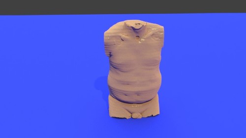
Chest wall - Skin model STL file from converted CT scan
By embodi3d
The chest wall (thoracic cage) is composed by twelve pairs of ribs laterally and the sternum anteriorly. The ribs are attached to the dorsal vertebrae (thoracic spine) posteriorly and along their costal cartilage to the sternum.
The thoracic cage main function is to protect the vital chest organs such as the heart and lungs. There are five muscles that make up the thoracic cage; the intercostals (external, internal and innermost), subcostals and transversus thoracis. This 3D model was created from the file STS_040 for a 57 years old female with breast implants. The original CT examination can be reviewed at: The 3D bone model created from this scan can be reviewed at: The 3D muscle model created from this scan can be reviewed at:9 downloads
- chest wall
- thoracic cage
- (and 11 more)
(0 reviews)0 comments
Updated
-

Free
(0 reviews)0 comments
Updated
-
Free
Whole Body - Skin model STL file from converted CT scan
By embodi3d
Whole body: chest, abdomen and pelvis
The chest wall (thoracic cage) is composed by twelve pairs of ribs laterally and the sternum anteriorly. The ribs are attached to the dorsal vertebrae (thoracic spine) posteriorly and along their costal cartilage to the sternum. The thoracic cage main function is to protect the vital chest organs such as the heart and lungs. The cervical spine is the upper most spines forming the spinal column, extending from the skull base to the level of the thoracic vertebra (the spines with attached ribs). The cervical spines are usually seven and the main function is to support the skull and to protect the spinal cord. The dorsal (thoracic) spine forms the middle portion of the vertebral column extending below the seventh cervical vertebra to above the first lumbar vertebra. The dorsal spine is formed by twelve vertebral bodies.
The vertebrae forming the dorsal spine are unique in shape as they are the only vertebral bodies articulating with ribs. The lumbar spine represents the mid-lower segment of the vertebral column and is composed of five adjacent vertebrae. They are convex anteriorly to form a lumbar lordosis. The lumbar spine facet joints allows limited movements and rotation. The bony pelvis is formed by 4 bones; a pair of hip bones, the sacrum and the coccyx. The bony pelvis supports the pelvic viscera and works to transmit force from the axial skeleton to the lower limbs.
The two hip bones are related anteriorly by the symphysis pubis and posteriorly to the sacroiliac joints bilaterally.
The original CT examination can be reviewed at:
The 3D bone model created from this scan can be reviewed at: The 3D muscle model created from this scan can be reviewed at:10 downloads
(0 reviews)0 comments
Updated
-

Free
Pelvis and Hip - Skin model STL file from converted CT scan
By embodi3d
The bony pelvis is formed by 4 bones; a pair of hip bones, the sacrum and the coccyx. The bony pelvis supports the pelvic viscera and works to transmit force from the axial skeleton to the lower limbs.
The two hip bones are related anteriorly by the symphysis pubis and posteriorly to the sacroiliac joints bilaterally.
The hip joint is a large synovial socket and ball joint which is formed by the femoral head (the ball) and the acetabulum (the socket). The acetabulum is formed by pelvic bones; the ilium, the ischium and the pubis. The hip joint represents the articulation between the lower extremity and the axial skeleton and allows a high degree of mobility while being stable. This model shows parts of the fingers as the patient's hands were set beside the body. The CT scan is derived from the file STS_037 The original CT examination can be reviewed at: The 3D bone model created from this scan can be reviewed at: The 3D muscle model created from this scan can be reviewed at:1 download
(0 reviews)0 comments
Updated
-

Free
Left Shoulder - Skin model STL file from converted CT scan
By embodi3d
The shoulder joint is a large and complex ball and socket joint formed by the humerus and the scapula (glenohumeral joint) while the clavicle join the acromion to form the acromioclavicular joint.
The shoulder joint is the most mobile joint in the human body on cost of instability. Lot of elements share to compensate the instability such as rotator cuff muscles, tendons and ligaments as well as the glenoid labrum. Muscles of the shoulder joint
The rotator cuff: supraspinatus, infraspinatus, teres minor and subscapularis
Posterior muscle group: deltoid, latissimus dorsi and teres major
Anterior muscle group: pectoralis major and coracobrachialis This 3D model was created from the file STS_037 The original CT examination can be reviewed at: The 3D bone model created from this scan can be reviewed at: The 3D muscle model created from this scan can be reviewed at:6 downloads
- shoulder joint
- ball and socket joint
- (and 13 more)
(1 review)0 comments
Updated
-

Free
Right Shoulder - undifferentiated pleomorphic spindle cell sarcoma - Skin Model STL file from converted CT scan
By embodi3d
This 3D model represents a case of undifferentiated pleomorphic spindle cell sarcoma implicating the right parascapular region of a 61 years old male. The patient represented with lung metastasis and was treated by surgical excision follower by chemotherapy as well as radiotherapy.
A cross sectional CT image is attached showing the lesion in axial, coronal and sagittal planes. Unfortunately pleomorphic undifferentiated sarcoma has an aggressive biological behaviour and a poor prognosis. Pleomorphic undifferentiated sarcomas can occur almost anywhere in the body, they have a predilection for the retroperitoneum and proximal extremities. They are usually confined to the soft tissues, but occasionally may arise in or from bone. This 3D model was created from the file STS_037 The original CT examination can be reviewed at: The 3D bone model created from this scan can be reviewed at: The 3D muscle model created from this scan can be reviewed at:
1 download
- shoulder
- spindle cell sarcoma
- (and 8 more)
(0 reviews)0 comments
Updated
-

Free
Chest wall skin model - STL file from converted CT scan
By embodi3d
The chest wall (thoracic cage) is composed by twelve pairs of ribs laterally and the sternum anteriorly. The ribs are attached to the dorsal vertebrae (thoracic spine) posteriorly and along their costal cartilage to the sternum.
The thoracic cage main function is to protect the vital chest organs such as the heart and lungs. There are five muscles that make up the thoracic cage; the intercostals (external, internal and innermost), subcostals, and transversus thoracis. This 3D model was created from the file STS_036 The original CT examination can be reviewed at: The 3D bone model created from this scan can be reviewed at: The 3D muscle model created from this scan can be reviewed at:3 downloads
- chest wall
- thoracic cage
- (and 12 more)
(0 reviews)0 comments
Updated
-

Free
(0 reviews)0 comments
Updated
-

Free
(0 reviews)0 comments
Submitted
-

Free
(0 reviews)0 comments
Updated
-

Free
test fiducial crane - stl file processed
By yas
test fiducial crane - stl file processed
0 downloads
(0 reviews)0 comments
Updated
-

Free
(0 reviews)0 comments
Submitted


-
File Reviews
-
File Comments
-
Recent Forum Posts
-

By jangiddrrk · Posted
Free connections, find your partner with no obligations Verified Damsels [URL=https://datesnow.life]Outstanding Сasual Dating[/URL] -
 Hello everyone, I hope this message finds you well. I am reaching out to our community in hopes of finding assistance with a project I'm currently undertaking. Specifically, I am in need of a detailed 3D model of the temporomandibular joint (TMJ). If anyone in our community has access to or expertise in creating high-quality 3D models, particularly of the temporomandibular joint, I would greatly appreciate any assistance you can offer. Whether you have a model readily available or can provide guidance on where to find one, your contribution would be invaluable to my project. Thank you in advance for your assistance and support.
 -

-

-

-


