
-
Welcome to embodi3D Downloads! This is the largest and fastest growing library of 3D printable anatomic models generated from real medical scans on the Internet. A unique scientific resource, most of the material is free. Registered members can download, upload, and sell models. To convert your own medical scans to a 3D model, take a look at democratiz3D, our free and automated conversion service.
Alert (6/17/22) - The democratiz3D scan-to-model conversion app is down due to a technical issue. We are working on a solution.
Skin
Models of the skin and body surface
773 files
-

Free
Pelvis - Skin model STL file from converted CT scan
The bony pelvis is formed by 4 bones; a pair of hip bones, the sacrum and the coccyx. The bony pelvis supports the pelvic viscera and works to transmit force from the axial skeleton to the lower limbs.
The two hip bones are related anteriorly by the symphysis pubis and posteriorly to the sacroiliac joints bilaterally. This model shows some irregular shaped pieces related to the contrast media within the colon as well as the femoral arteries. This 3D model was created from the file ABD_LYMPH_001 The original CT examination can be reviewed at: The 3D bone model created from this scan can be reviewed at: The 3D muscle model created from this scan can be reviewed at:0 downloads
(0 reviews)0 comments
Updated
-

Free
(0 reviews)0 comments
Submitted
-
Free
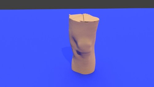
Left knee - Skin model STL file from converted CT scan
By embodi3d
The knee joint is formed by three bones: the femur, the tibia and the patella. the knee joint is the largest synovial joint and provides the flexion and extension movements of the leg as well as relative medial and lateral rotations while in relative flexion.
The knee joint articulations are two condylar joints between the femur and the tibia as well as a joint between the patella and the femur. Although the fibula is closely related to the knee joint but it doesn't share in articulation. The knee joint is also formed by some ligaments and cartilage called (menisci) which are best imaged by MRI. This 3D model was created from the file STS_051 The original CT examination can be reviewed at: The 3D bone model created from this scan can be reviewed at: The 3D muscle model created from this scan can be reviewed at:28 downloads
(0 reviews)0 comments
Updated
-

Free
Left Ankle - Skin model STL file from converted CT scan
By embodi3d
The ankle joint is comprised of the tibia, fibula, talus, and calcaneus as well as the supporting ligaments, muscles and neurovascular bundles.
The ankle is a synovial joint composed of the distal tibia and fibula as they articulate with the talus. The distal tibia and fibula articulate with each other at the distal tibiofibular joint which is more commonly referred to as the tibiofibular syndesmosis. This 3D model was created from the file STS_051 The original CT examination can be reviewed at: The 3D bone model created from this scan can be reviewed at: The 3D muscle model created from this scan can be reviewed at:4 downloads
(0 reviews)0 comments
Updated
-

Free
Right knee - Skin model STL file from converted CT scan
By embodi3d
The knee joint is formed by three bones: the femur, the tibia and the patella. the knee joint is the largest synovial joint and provides the flexion and extension movements of the leg as well as relative medial and lateral rotations while in relative flexion.
The knee joint articulations are two condylar joints between the femur and the tibia as well as a joint between the patella and the femur. Although the fibula is closely related to the knee joint but it doesn't share in articulation. The knee joint is also formed by some ligaments and cartilage called (menisci) which are best imaged by MRI. This 3D model was created from the file STS_051 The original CT examination can be reviewed at: The 3D bone model created from this scan can be reviewed at: The 3D muscle model created from this scan can be reviewed at:141 downloads
(0 reviews)0 comments
Updated
-

Free
Right Ankle - Skin model STL file from converted CT scan
By embodi3d
The ankle joint is comprised of the tibia, fibula, talus, and calcaneus as well as the supporting ligaments, muscles and neurovascular bundles.
The ankle is a synovial joint composed of the distal tibia and fibula as they articulate with the talus. The distal tibia and fibula articulate with each other at the distal tibiofibular joint which is more commonly referred to as the tibiofibular syndesmosis. This 3D model was created from the file STS_051 The original CT examination can be reviewed at: The 3D bone model created from this scan can be reviewed at: The 3D muscle model created from this scan can be reviewed at:1 download
- ankle joint
- tibia
- (and 11 more)
(0 reviews)0 comments
Updated
-

Free
Pelvis and Hip - Skin model STL file from converted CT scan
By embodi3d
The bony pelvis is formed by 4 bones; a pair of hip bones, the sacrum and the coccyx. The bony pelvis supports the pelvic viscera and works to transmit force from the axial skeleton to the lower limbs.
The two hip bones are related anteriorly by the symphysis pubis and posteriorly to the sacroiliac joints bilaterally. The hip joint is a large synovial socket and ball joint which is formed by the femoral head (the ball) and the acetabulum (the socket). The acetabulum is formed by pelvic bones; the ilium, the ischium and the pubis. The hip joint represents the articulation between the lower extremity and the axial skeleton and allows a high degree of mobility while being stable. This 3D model was created from the file STS_040 The original CT examination can be reviewed at: The 3D bone model created from this scan can be reviewed at: The 3D muscle model created from this scan can be reviewed at:20 downloads
(0 reviews)0 comments
Updated
-

Free
Whole Body - Skin model STL file from converted CT scan
By embodi3d
Whole body: chest, abdomen and pelvis
The chest wall (thoracic cage) is composed by twelve pairs of ribs laterally and the sternum anteriorly. The ribs are attached to the dorsal vertebrae (thoracic spine) posteriorly and along their costal cartilage to the sternum.
The thoracic cage main function is to protect the vital chest organs such as the heart and lungs.
The cervical spine is the upper most spines forming the spinal column, extending from the skull base to the level of the thoracic vertebra (the spines with attached ribs). The cervical spines are usually seven and the main function is to support the skull and to protect the spinal cord.
The dorsal (thoracic) spine forms the middle portion of the vertebral column extending below the seventh cervical vertebra to above the first lumbar vertebra. The dorsal spine is formed by twelve vertebral bodies.
The vertebrae forming the dorsal spine are unique in shape as they are the only vertebral bodies articulating with ribs.
The lumbar spine represents the mid-lower segment of the vertebral column and is composed of five adjacent vertebrae. They are convex anteriorly to form a lumbar lordosis. The lumbar spine facet joints allows limited movements and rotation.
The bony pelvis is formed by 4 bones; a pair of hip bones, the sacrum and the coccyx. The bony pelvis supports the pelvic viscera and works to transmit force from the axial skeleton to the lower limbs.
The two hip bones are related anteriorly by the symphysis pubis and posteriorly to the sacroiliac joints bilaterally.
This 3D model was created from the file STS_040
The original CT examination can be reviewed at: The 3D bone model created from this scan can be reviewed at: The 3D muscle model created from this scan can be reviewed at:24 downloads
(0 reviews)0 comments
Updated
-

Free
Scan Septembre 2017 - stl file processed
By kelu28
Scan Septembre 2017 - stl file processed1 download
(0 reviews)0 comments
Submitted
-

Free
Scan Septembre 2017 - stl file processed
By kelu28
Scan Septembre 2017 - stl file processed1 download
(0 reviews)0 comments
Submitted
-

Free
Scan Septembre 2017 - stl file processed
By kelu28
Scan Septembre 2017 - stl file processed1 download
(0 reviews)0 comments
Submitted
-

Free
Scan Septembre 2017 - stl file processed
By kelu28
Scan Septembre 2017 - stl file processed1 download
(0 reviews)0 comments
Submitted
-

Free
Scan Septembre 2017 - stl file processed
By kelu28
Scan Septembre 2017 - stl file processed1 download
(0 reviews)0 comments
Submitted
-

Free
(0 reviews)0 comments
Submitted
-

Free
(0 reviews)0 comments
Updated
-

Free
(0 reviews)0 comments
Updated
-

Free
75 - stl file processed
By Mike_0983
75 - stl file processed
1 download
- knee
- lower limb
- (and 3 more)
(0 reviews)0 comments
Updated
-

Free
(0 reviews)0 comments
Updated
-

Free
200 - stl file processed
By Mike_0983
200 - stl file processed
1 download
- knee
- lower limb
- (and 3 more)
(0 reviews)0 comments
Updated
-

Free
(0 reviews)0 comments
Updated
-

Free
(0 reviews)0 comments
Updated
-

Free
(0 reviews)0 comments
Updated
-

Free
(0 reviews)0 comments
Submitted
-

Free
(0 reviews)0 comments
Updated
-

Free
(0 reviews)0 comments
Updated


-
File Reviews
-
File Comments
-
Recent Forum Posts
-

By jangiddrrk · Posted
Free connections, find your partner with no obligations Verified Damsels [URL=https://datesnow.life]Outstanding Сasual Dating[/URL] -
 Hello everyone, I hope this message finds you well. I am reaching out to our community in hopes of finding assistance with a project I'm currently undertaking. Specifically, I am in need of a detailed 3D model of the temporomandibular joint (TMJ). If anyone in our community has access to or expertise in creating high-quality 3D models, particularly of the temporomandibular joint, I would greatly appreciate any assistance you can offer. Whether you have a model readily available or can provide guidance on where to find one, your contribution would be invaluable to my project. Thank you in advance for your assistance and support.
 -

-

-

-


