
-
Welcome to embodi3D Downloads! This is the largest and fastest growing library of 3D printable anatomic models generated from real medical scans on the Internet. A unique scientific resource, most of the material is free. Registered members can download, upload, and sell models. To convert your own medical scans to a 3D model, take a look at democratiz3D, our free and automated conversion service.
Alert (6/17/22) - The democratiz3D scan-to-model conversion app is down due to a technical issue. We are working on a solution.
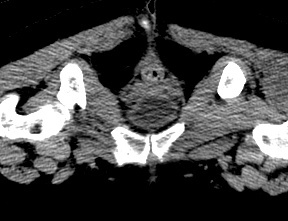
Abdomen and Pelvis CTs
CT scans of the abdomen and pelvis
674 files
-
Free
Pelvic Bones (female pelvis) - CT Scan
By embodi3d
The bony pelvis is formed by 4 bones; a pair of hip bones, the sacrum and the coccyx. The bony pelvis supports the pelvic viscera and works to transmit force from the axial skeleton to the lower limbs. The two hip bones are related anteriorly by the symphysis pubis and posteriorly to the sacroiliac joints bilaterally. This model if for a 57 years old female pelvis, it shows some irregular shaped pieces related to the contrast media within the colon as well as the upper halves of the femoral bones. The CT scan is derived from the file STS_040 The 3D bone model created from this scan can be reviewed at: The 3D muscle model created from this scan can be reviewed at:42 downloads
(0 reviews)0 comments
Updated
-

Free
Pelvis and Hip - CT scan
By embodi3d
The bony pelvis is formed by 4 bones; a pair of hip bones, the sacrum and the coccyx. The bony pelvis supports the pelvic viscera and works to transmit force from the axial skeleton to the lower limbs.
The two hip bones are related anteriorly by the symphysis pubis and posteriorly to the sacroiliac joints bilaterally. The hip joint is a large synovial socket and ball joint which is formed by the femoral head (the ball) and the acetabulum (the socket). The acetabulum is formed by pelvic bones; the ilium, the ischium and the pubis. The hip joint represents the articulation between the lower extremity and the axial skeleton and allows a high degree of mobility while being stable. The CT scan is derived from the file STS_037 The 3D bone model created from this scan can be reviewed at: The 3D muscle model created from this scan can be reviewed at: The 3D skin model created from this scan can be reviewed at:22 downloads
(0 reviews)0 comments
Updated
-

Free
CT Scan Conversion Tutorial
By Mdoddo
Chest Abdomen Pelvis CT to covert to 3d printable file
CT without contrast. abdomen pelvis, stl, axial, dicom, lung, liver
4 downloads
- ct without contrast
- abdomen
- (and 6 more)
(0 reviews)0 comments
Updated
-

Free
(0 reviews)0 comments
Updated
-

Free
(0 reviews)0 comments
Updated
-

Free
Sacrum CT scan
By embodi3d
The sacrum is the lower most segment of the vertebral column and also forms the posterior wall of the bony pelvis. The sacrum is formed by five fused sacral vertebrae. The sacrum is formed by fusion of five sacral vertebrae has three surfaces, a base and an apex. The body of the first segment is large and is similar to lumbar vertebra whereas the bodies of the next bones get progressively smaller, are flattened from the back, and curved to shape. The sacrum articulates with four other bones – iliac bones on either side, L5 above and coccyx below. It is tilted forward and curved with anterior concavity and posterior convexity allowing greater room for pelvic cavity. The curvature of sacrum varies in individuals. This CT scan was cropped from the file STS_036 The 3D model created from this scan can be reviewed at:1 download
- sacrum
- vertebral column
- (and 6 more)
(0 reviews)0 comments
Updated
-

Free
Female Pelvic Bones CT Scan
By embodi3d
Pelvic Bones 3D Printable STL File Converted From CT Scan This 3D model was created from the file STS_036. A 3D printable STL file model created from this scan can be found here.
37 downloads
(0 reviews)0 comments
Updated
-
(0 reviews)
0 comments
Submitted
-
(0 reviews)
0 comments
Submitted
-

Free
Pelvic Bones 3D Printable STL File Converted From CT Scan
By embodi3d
Pelvic Bones 3D Printable STL File Converted From CT Scan
A 3D printable STL file model created from this scan can be found here.
11 downloads
(0 reviews)0 comments
Updated
-

Free
(0 reviews)0 comments
Updated
-

Free
(0 reviews)0 comments
Submitted
-

Free
(0 reviews)0 comments
Updated
-
(0 reviews)
0 comments
Updated
-

Free
Left Kidney with Tumor
By kminars
This is a scan of a left kidney with a tumor.
kidney, tumor, ct with contrast, abdominal, .stl, bowel, CTA, liver, spleen, great, vessels, porta, vein, small, bowel, psoas, muscle
50 downloads
(0 reviews)0 comments
Updated
-

Free
(0 reviews)0 comments
Submitted
-

Free
(0 reviews)0 comments
Submitted
-

Free
(0 reviews)0 comments
Submitted
-

Free
(0 reviews)0 comments
Submitted
-

Free
(0 reviews)0 comments
Submitted
-

Free
ABDOMEN
By Vishwas
This is an Abdomen Dual Phase File
dicom, stl, axial, abdomen, pelvis, liver, spleen, vertebrae,
10 downloads
(0 reviews)0 comments
Updated
-

Free
(0 reviews)0 comments
Submitted
-

Free
(0 reviews)0 comments
Submitted
-

Free
(0 reviews)0 comments
Submitted
-

Free
(0 reviews)0 comments
Submitted


-
File Reviews
-
File Comments
-
Recent Forum Posts
-

By jangiddrrk · Posted
Free connections, find your partner with no obligations Verified Damsels [URL=https://datesnow.life]Outstanding Сasual Dating[/URL] -
 Hello everyone, I hope this message finds you well. I am reaching out to our community in hopes of finding assistance with a project I'm currently undertaking. Specifically, I am in need of a detailed 3D model of the temporomandibular joint (TMJ). If anyone in our community has access to or expertise in creating high-quality 3D models, particularly of the temporomandibular joint, I would greatly appreciate any assistance you can offer. Whether you have a model readily available or can provide guidance on where to find one, your contribution would be invaluable to my project. Thank you in advance for your assistance and support.
 -

-

-

-


