


About This File
The bony pelvis is formed by 4 bones; a pair of hip bones, the sacrum and the coccyx. The bony pelvis supports the pelvic viscera and works to transmit force from the axial skeleton to the lower limbs.
The two hip bones are related anteriorly by the symphysis pubis and posteriorly to the sacroiliac joints bilaterally.
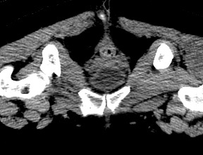
This model if for a 57 years old female pelvis, it shows some irregular shaped pieces related to the contrast media within the colon as well as the upper halves of the femoral bones.
The CT scan is derived from the file STS_040
The 3D bone model created from this scan can be reviewed at:
The 3D muscle model created from this scan can be reviewed at:


