
-
Welcome to embodi3D Downloads! This is the largest and fastest growing library of 3D printable anatomic models generated from real medical scans on the Internet. A unique scientific resource, most of the material is free. Registered members can download, upload, and sell models. To convert your own medical scans to a 3D model, take a look at democratiz3D, our free and automated conversion service.
Alert (6/17/22) - The democratiz3D scan-to-model conversion app is down due to a technical issue. We are working on a solution.
Spine CTs
CTs of the spine
319 files
-

Free
(0 reviews)0 comments
Updated
-

Free
Spine Test 10:59 10/10/2017 edited at 11:07
By embodi3d
Test first spine at 10:59 on 10/10
Edited spine test at 11:07 on 10/10
0 downloads
(0 reviews)0 comments
Updated
-
CTScan_SpinalCordStimulator
By jjfranzen
A CT scan of my lumber region that shows my SCS and the wires going along my spinal column.
2 downloads
(0 reviews)0 comments
Submitted
-

Free
(0 reviews)0 comments
Submitted
-

Free
Lumbo-Sacral Spine - CT scan
By embodi3d
The lumbar spine represents the mid-lower segment of the vertebral column and is composed of five adjacent vertebrae. They are convex anteriorly to form a lumbar lordosis. The lumbar spine facet joints allows limited movements and rotation. Each lumbar vertebra is formed of: A body which is kidney shaped and is convex anteriorly while flattened posteriorly, pedicles and lamina, transverse processes, articular processes and a spinous process.
The sacrum is the lower most segment of the vertebral column and also forms the posterior wall of the bony pelvis. The sacrum is formed by five fused sacral vertebrae. The sacrum is formed by fusion of five sacral vertebrae has three surfaces, a base and an apex. The body of the first segment is large and is similar to lumbar vertebra whereas the bodies of the next bones get progressively smaller, are flattened from the back, and curved to shape. The sacrum articulates with four other bones – iliac bones on either side, L5 above and coccyx below. It is tilted forward and curved with anterior concavity and posterior convexity allowing greater room for pelvic cavity. The curvature of sacrum varies in individuals. The CT scan is derived from the file STS_040 The 3D bone model created from this scan can be reviewed at:5 downloads
- lumbar spine
- sacral spine
- (and 14 more)
(0 reviews)0 comments
Updated
-

Free
Lumbar Spine - CT scan
By embodi3d
The lumbar spine represents the mid-lower segment of the vertebral column and is composed of five adjacent vertebrae. They are convex anteriorly to form a lumbar lordosis. The lumbar spine facet joints allows limited movements and rotation. Each lumbar vertebra is formed of: A body which is kidney shaped and is convex anteriorly while flattened posteriorly, pedicles and lamina, transverse processes, articular processes and a spinous process. The CT scan is derived from the file STS_040 The 3D bone model created from this scan can be reviewed at:17 downloads
- lumbar spine
- lumbar vertebra
- (and 9 more)
(0 reviews)0 comments
Updated
-

Free
Dorsal Spine - CT scan
By embodi3d
The dorsal (thoracic) spine forms the middle portion of the vertebral column extending below the seventh cervical vertebra to above the first lumbar vertebra. The dorsal spine is formed by twelve vertebral bodies.
The vertebrae forming the dorsal spine are unique in shape as they are the only vertebral bodies articulating with ribs. The CT scan is derived from the file STS_040 The 3D bone model created from this scan can be reviewed at:7 downloads
- dorsal spine
- thoracic spine
- (and 8 more)
(0 reviews)0 comments
Updated
-

Free
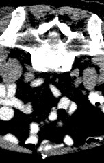
Cervical Spine - CT scan
By embodi3d
The cervical spine is the upper most spines forming the spinal column, extending from the skull base to the level of the thoracic vertebra (the spines with attached ribs). The cervical spines are usually seven and the main function is to support the skull and to protect the spinal cord. Apart from the first cervical vertebra (atlas) and the second vertebra (axis), the other vertebral bodies share a general anatomical appearance:
Oval shaped vertebral bodies with wide vertebral arch, large vertebral foramina and long spinous processes. The CT scan is derived from the file STS_040 The 3D bone model created from this scan can be reviewed at:27 downloads
- cervical spine
- cervical vertebra
- (and 6 more)
(0 reviews)0 comments
Updated
-
(0 reviews)
0 comments
Submitted
-

Free
Whole Spine (Cervical-Dorsal-Lumbar-Sacral) - CT scan
By embodi3d
Whole Spine (Cervical-Dorsal-Lumbar-Sacral) The cervical spine is the upper most spines forming the spinal column, extending from the skull base to the level of the thoracic vertebra (the spines with attached ribs). The cervical spines are usually seven and the main function is to support the skull and to protect the spinal cord.
Apart from the first cervical vertebra (atlas) and the second vertebra (axis), the other vertebral bodies share a general anatomical appearance:
Oval shaped vertebral bodies with wide vertebral arch, large vertebral foramina and long spinous processes.
The dorsal (thoracic) spine forms the middle portion of the vertebral column extending below the seventh cervical vertebra to above the first lumbar vertebra. The dorsal spine is formed by twelve vertebral bodies.
The vertebrae forming the dorsal spine are unique in shape as they are the only vertebral bodies articulating with ribs.
The lumbar spine represents the mid-lower segment of the vertebral column and is composed of five adjacent vertebrae. They are convex anteriorly to form a lumbar lordosis. The lumbar spine facet joints allows limited movements and rotation.
This model shows lumbar spondylo-degenerative changes manifested by marginal osteophytic lipping.
The sacrum is the lower most segment of the vertebral column and also forms the posterior wall of the bony pelvis. The sacrum is formed by five fused sacral vertebrae.
The sacrum is formed by fusion of five sacral vertebrae has three surfaces, a base and an apex. The body of the first segment is large and is similar to lumbar vertebra whereas the bodies of the next bones get progressively smaller, are flattened from the back, and curved to shape.
The sacrum articulates with four other bones – iliac bones on either side, L5 above and coccyx below. It is tilted forward and curved with anterior concavity and posterior convexity allowing greater room for pelvic cavity. The curvature of sacrum varies in individuals. The CT scan is derived from the file STS_040 The 3D bone model created from this scan can be reviewed at:54 downloads
- cervical spine
- dorsal spine
- (and 10 more)
(0 reviews)0 comments
Updated
-

Free
(0 reviews)0 comments
Updated
-

Free
L5 vertebra - CT scan
By embodi3d
Each lumbar vertebra is formed of: A body which is kidney shaped and is convex anteriorly while flattened posteriorly, pedicles and lamina, transverse processes, articular processes and a spinous process. This model shows the whole vertebral body with related articulations of the lower level vertebra. The CT scan is derived from the file STS_037 The 3D bone model created from this scan can be reviewed at:4 downloads
- lumbar spine
- lumbar vertebra
- (and 7 more)
(0 reviews)0 comments
Updated
-

Free
L4 vertebra - CT scan
By embodi3d
Each lumbar vertebra is formed of: A body which is kidney shaped and is convex anteriorly while flattened posteriorly, pedicles and lamina, transverse processes, articular processes and a spinous process. This model shows the whole vertebral body with related articulations of the lower level vertebra. The CT scan is derived from the file STS_037 The 3D bone model created from this scan can be reviewed at:5 downloads
- lumbar spine
- lumbar vertebra
- (and 7 more)
(0 reviews)0 comments
Updated
-

Free
L3 vertebra - CT scan
By embodi3d
Each lumbar vertebra is formed of: A body which is kidney shaped and is convex anteriorly while flattened posteriorly, pedicles and lamina, transverse processes, articular processes and a spinous process. This model shows the whole vertebral body with related articulations of the lower level vertebra. The CT scan is derived from the file STS_037 The 3D bone model created from this scan can be reviewed at:4 downloads
- lumbar spine
- lumbar vertebra
- (and 7 more)
(0 reviews)0 comments
Updated
-

Free
L2 vertebra - CT scan
By embodi3d
Each lumbar vertebra is formed of: A body which is kidney shaped and is convex anteriorly while flattened posteriorly, pedicles and lamina, transverse processes, articular processes and a spinous process. This model shows the whole vertebral body with related articulations of the lower level vertebra. The CT scan is derived from the file STS_037 The 3D bone model created from this scan can be reviewed at:1 download
- lumbar vertebra
- lumbar spine
- (and 7 more)
(0 reviews)0 comments
Updated
-

Free
L1 vertebra - CT scan
By embodi3d
Each lumbar vertebra is formed of: A body which is kidney shaped and is convex anteriorly while flattened posteriorly, pedicles and lamina, transverse processes, articular processes and a spinous process. This model shows the whole vertebral body with related articulations of the lower level vertebra. The CT scan is derived from the file STS_037 The 3D bone model created from this scan can be reviewed at:3 downloads
- lumbar spine
- lumbar vertebra
- (and 7 more)
(0 reviews)0 comments
Updated
-
Free
Whole Spine (Cervical-Dorsal-Lumbar-Sacral) - CT scan
By embodi3d
The cervical spine is the upper most spines forming the spinal column, extending from the skull base to the level of the thoracic vertebra (the spines with attached ribs). The cervical spines are usually seven and the main function is to support the skull and to protect the spinal cord. Apart from the first cervical vertebra (atlas) and the second vertebra (axis), the other vertebral bodies share a general anatomical appearance:
Oval shaped vertebral bodies with wide vertebral arch, large vertebral foramina and long spinous processes. The dorsal (thoracic) spine forms the middle portion of the vertebral column extending below the seventh cervical vertebra to above the first lumbar vertebra. The dorsal spine is formed by twelve vertebral bodies.
The vertebrae forming the dorsal spine are unique in shape as they are the only vertebral bodies articulating with ribs. The lumbar spine represents the mid-lower segment of the vertebral column and is composed of five adjacent vertebrae. They are convex anteriorly to form a lumbar lordosis. The lumbar spine facet joints allows limited movements and rotation. The sacrum is the lower most segment of the vertebral column and also forms the posterior wall of the bony pelvis. The sacrum is formed by five fused sacral vertebrae. The sacrum is formed by fusion of five sacral vertebrae has three surfaces, a base and an apex. The body of the first segment is large and is similar to lumbar vertebra whereas the bodies of the next bones get progressively smaller, are flattened from the back, and curved to shape. The sacrum articulates with four other bones – iliac bones on either side, L5 above and coccyx below. It is tilted forward and curved with anterior concavity and posterior convexity allowing greater room for pelvic cavity. The curvature of sacrum varies in individuals. The CT scan is derived from the file STS_037 The 3D bone model created from this scan can be reviewed at:12 downloads
- vertebral column
- cervical spine
- (and 5 more)
(0 reviews)0 comments
Updated
-
Free
Lumbo-Sacral Spine - CT scan
By embodi3d
The lumbar spine represents the mid-lower segment of the vertebral column and is composed of five adjacent vertebrae. They are convex anteriorly to form a lumbar lordosis. The lumbar spine facet joints allows limited movements and rotation.
The sacrum is the lower most segment of the vertebral column and also forms the posterior wall of the bony pelvis. The sacrum is formed by five fused sacral vertebrae. The sacrum is formed by fusion of five sacral vertebrae has three surfaces, a base and an apex. The body of the first segment is large and is similar to lumbar vertebra whereas the bodies of the next bones get progressively smaller, are flattened from the back, and curved to shape. The CT scan is derived from the file STS_037 The 3D bone model created from this scan can be reviewed at:3 downloads
- lumbar spine
- sacral spine
- (and 6 more)
(0 reviews)0 comments
Updated
-
(0 reviews)
0 comments
Submitted
-

Free
Sacral Spine - CT scan
By embodi3d
The sacrum is the lower most segment of the vertebral column and also forms the posterior wall of the bony pelvis. The sacrum is formed by five fused sacral vertebrae. The sacrum is formed by fusion of five sacral vertebrae has three surfaces, a base and an apex. The body of the first segment is large and is similar to lumbar vertebra whereas the bodies of the next bones get progressively smaller, are flattened from the back, and curved to shape. The sacrum articulates with four other bones – iliac bones on either side, L5 above and coccyx below. It is tilted forward and curved with anterior concavity and posterior convexity allowing greater room for pelvic cavity. The curvature of sacrum varies in individuals. The CT scan is derived from the file STS_037 The 3D bone model created from this scan can be reviewed at:1 download
- sacral spine
- sacral vertebrae
- (and 2 more)
(0 reviews)0 comments
Updated
-

Free
Lumbar Spine - CT scan
By embodi3d
The lumbar spine represents the mid-lower segment of the vertebral column and is composed of five adjacent vertebrae. They are convex anteriorly to form a lumbar lordosis. The lumbar spine facet joints allows limited movements and rotation. Each lumbar vertebra is formed of: A body which is kidney shaped and is convex anteriorly while flattened posteriorly, pedicles and lamina, transverse processes, articular processes and a spinous process. The CT scan is derived from the file STS_037 The 3D bone model created from this scan can be reviewed at:9 downloads
- lumbar spine
- lumbar vertebrae
- (and 2 more)
(0 reviews)0 comments
Updated
-

Free
Dorsal Spine - CT scan
By embodi3d
The dorsal (thoracic) spine forms the middle portion of the vertebral column extending below the seventh cervical vertebra to above the first lumbar vertebra. The dorsal spine is formed by twelve vertebral bodies.
The vertebrae forming the dorsal spine are unique in shape as they are the only vertebral bodies articulating with ribs.
The CT scan is derived from the file STS_037
The 3D bone model created from this scan can be reviewed at:
5 downloads
- dorsal spine
- dorsal vertebrae
- (and 5 more)
(0 reviews)0 comments
Updated
-

Free
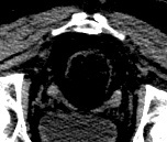
Cervical Spine - CT scan
By embodi3d
The cervical spine is the upper most spines forming the spinal column, extending from the skull base to the level of the thoracic vertebra (the spines with attached ribs). The cervical spines are usually seven and the main function is to support the skull and to protect the spinal cord. Apart from the first cervical vertebra (atlas) and the second vertebra (axis), the other vertebral bodies share a general anatomical appearance:
Oval shaped vertebral bodies with wide vertebral arch, large vertebral foramina and long spinous processes. The CT scan is derived from the file STS_037 The 3D bone model created from this scan can be reviewed at:11 downloads
- cervical spine
- cervical vertebrae
- (and 5 more)
(0 reviews)0 comments
Updated
-

Free
Dorsal Spine - CT scan
By embodi3d
The dorsal (thoracic) spine forms the middle portion of the vertebral column extending below the seventh cervical vertebra to above the first lumbar vertebra. The dorsal spine is formed by twelve vertebral bodies. The vertebrae forming the dorsal spine are unique in shape as they are the only vertebral bodies articulating with ribs. This scan is derived from the file STS_036 The 3D bone model created from this scan can be reviewed at:2 downloads
(0 reviews)0 comments
Updated
-

Free
Lumbar Spine - CT scan
By embodi3d
The lumbar spine represents the mid-lower segment of the vertebral column and is composed of five adjacent vertebrae. They are convex anteriorly to form a lumbar lordosis. The lumbar spine facet joints allows limited movements and rotation. Each lumbar vertebra is formed of: A body which is kidney shaped and is convex anteriorly while flattened posteriorly, pedicles and lamina, transverse processes, articular processes and a spinous process. This 3D model was created from the file STS_036 The 3D bone model created from this scan can be reviewed at:27 downloads
- lumbar spine
- lumbar vertebrae
- (and 9 more)
(0 reviews)0 comments
Updated


-
File Reviews
-
File Comments
-
Recent Forum Posts
-

By jangiddrrk · Posted
Free connections, find your partner with no obligations Verified Damsels [URL=https://datesnow.life]Outstanding Сasual Dating[/URL] -
 Hello everyone, I hope this message finds you well. I am reaching out to our community in hopes of finding assistance with a project I'm currently undertaking. Specifically, I am in need of a detailed 3D model of the temporomandibular joint (TMJ). If anyone in our community has access to or expertise in creating high-quality 3D models, particularly of the temporomandibular joint, I would greatly appreciate any assistance you can offer. Whether you have a model readily available or can provide guidance on where to find one, your contribution would be invaluable to my project. Thank you in advance for your assistance and support.
 -

-

-

-


