
Using 3D Printed Liver Models for Guidance During Liver Surgery

Entry posted by Dr. Mike ·
2,109 views

Researchers from Nagoya University in Japan are now using customized 3D printed liver models created from patient Computer Tomography (CT) scans for guidance during liver surgery, as reported at the 2014 Radiological Society of North America meeting. The human liver is a complex organ. Liver cells, called hepatocytes, do the work of cleaning the blood of toxins and waste -- the primary function of the liver. Hepatocytes are dependent on a complex network of vascular structures, including bile ducts, hepatic arteries, hepatic veins, and portal veins, which are organized into a complex branching network. This network is in turn organized into self contained units, each with its own bile ducts, arteries, and hepatic and portal veins, called segments, of which there are eight in the typical human liver.
When liver surgeons resect, or cut out, cancer from the liver, they remove the entire segment or segments that are involved, working hard to avoid any damage to segments that are uninvolved. The procedure is delicate, and there is no room for error. Cut out too little and there may be tumor left in the liver which can subsequently spread. Cut too much and a host of complications can arise, including bleeding, bile leak, liver failure, and potentially death. Furthermore, the surgeon must work under conditions where the tumor and vital structures may be poorly visualized or obscured by blood in the operative field.
This is where the 3D printed models come in. The Japanese researchers took CT scan data from the patient undergoing the liver cancer surgery and created a 3D printed model of the entire liver using a transparent material. They left the vascular structures hollow, and subsequently filled them with a colored material to color-code each type of vessel. The model was then vacuum sealed in a sterile pouch and brought unto the operating room during the surgery. The surgeon could handle the 3D model during the operation. Before making each cut, the surgeon could refer to the model and confirm where the tumor and each vital vessel was located, thus avoiding mistakes and reducing operating time.
Creating 3D printed models prior to surgery for preoperative planning and intraoperative guidance is something that I have done personally for endovascular procedures (a publication on my experience is forthcoming). I can tell you that the certainty of knowing what you are getting into, and avoiding nasty and unexpected surprises in the OR, is invaluable. Once surgeons experience the comfort of having an anatomically accurate intraoperative 3D printed reference model they will never want to go back. 3D printing in medicine is here to stay.
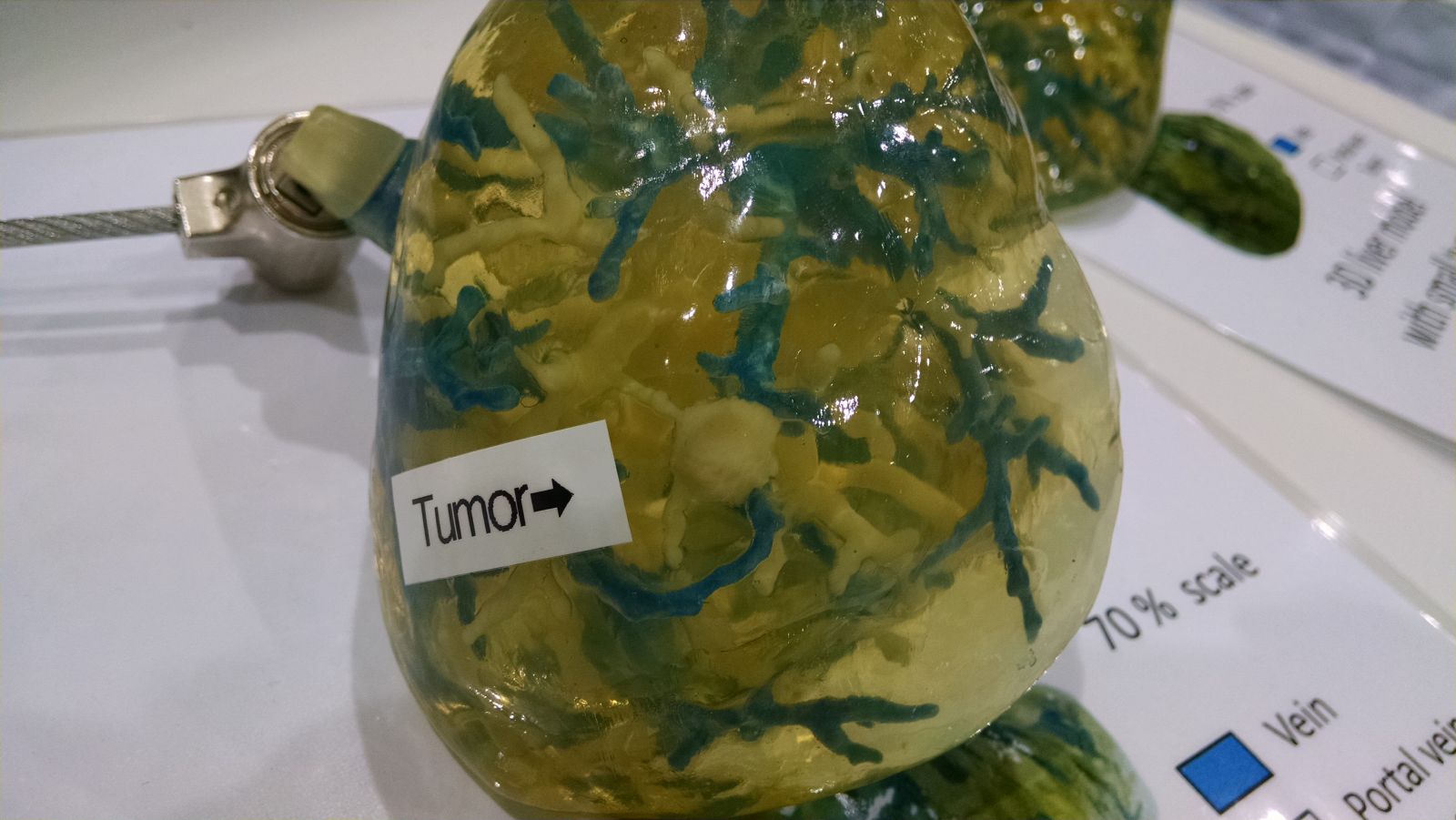
3D printed liver model showing the tumor to be resected.
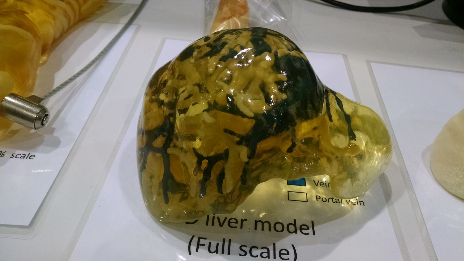
3D printed liver model
I will be publishing more blog articles about 3D printing at the 2014 RSNA meeting. Follow this blog or follow on Twitter @Embodi3D.


0 Comments
Recommended Comments
There are no comments to display.
Create an account or sign in to comment
You need to be a member in order to leave a comment
Create an account
Sign up for a new account in our community. It's easy!
Register a new accountSign in
Already have an account? Sign in here.
Sign In Now