
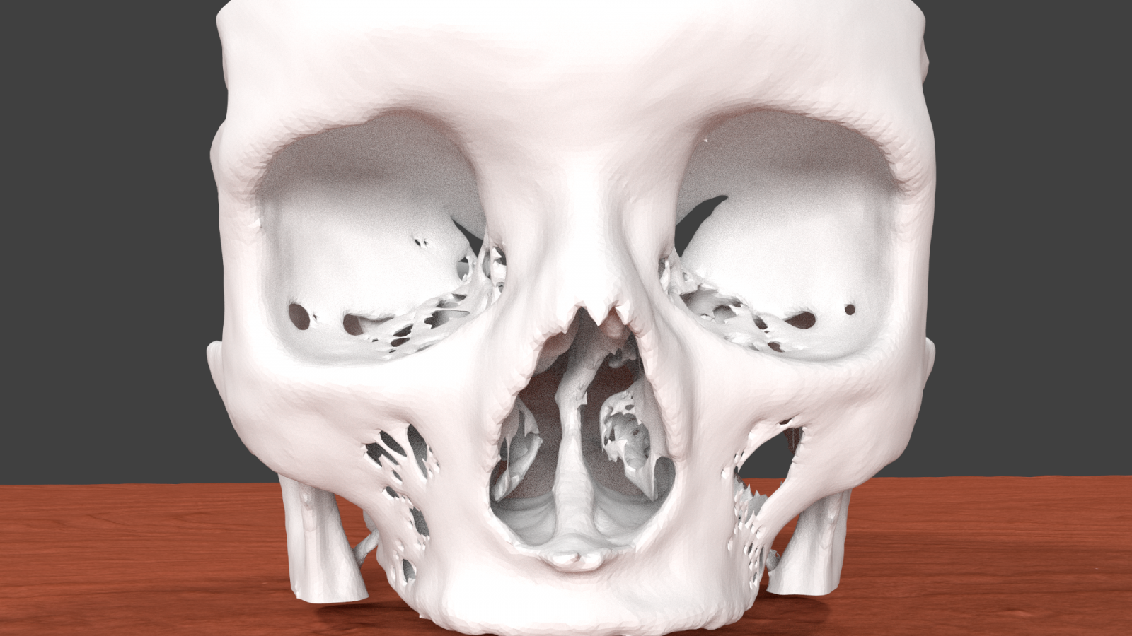
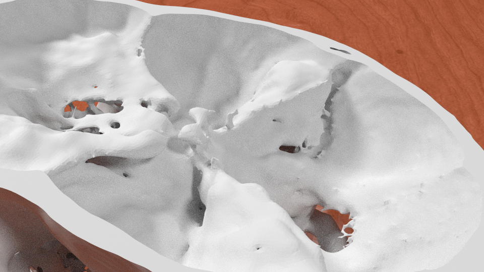
3D printed skull base generated from CT scan data accurately demonstrates complex skull base anatomy.

Entry posted by Dr. Mike ·
20,948 views
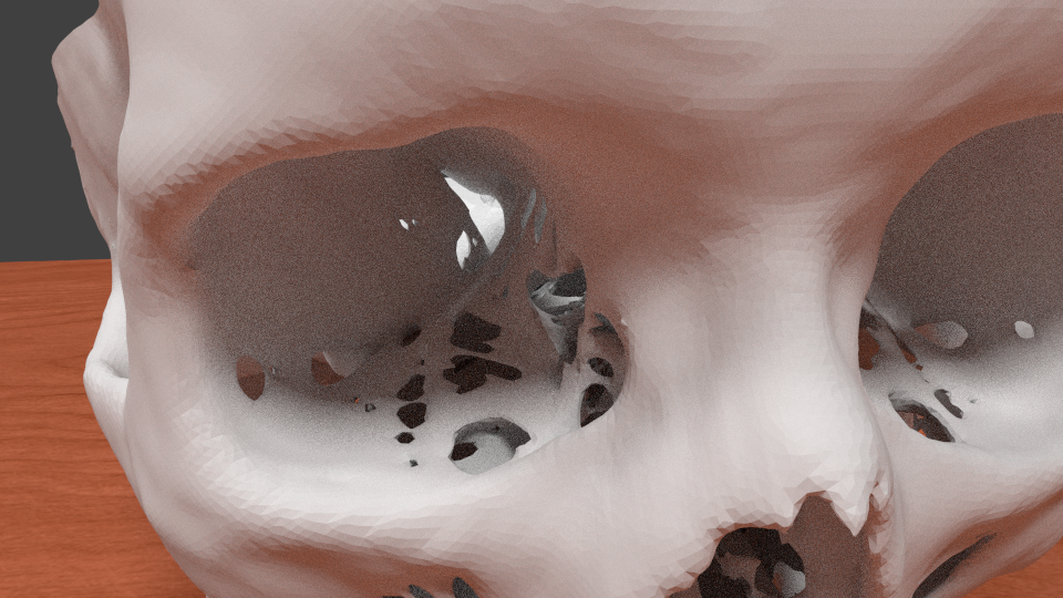
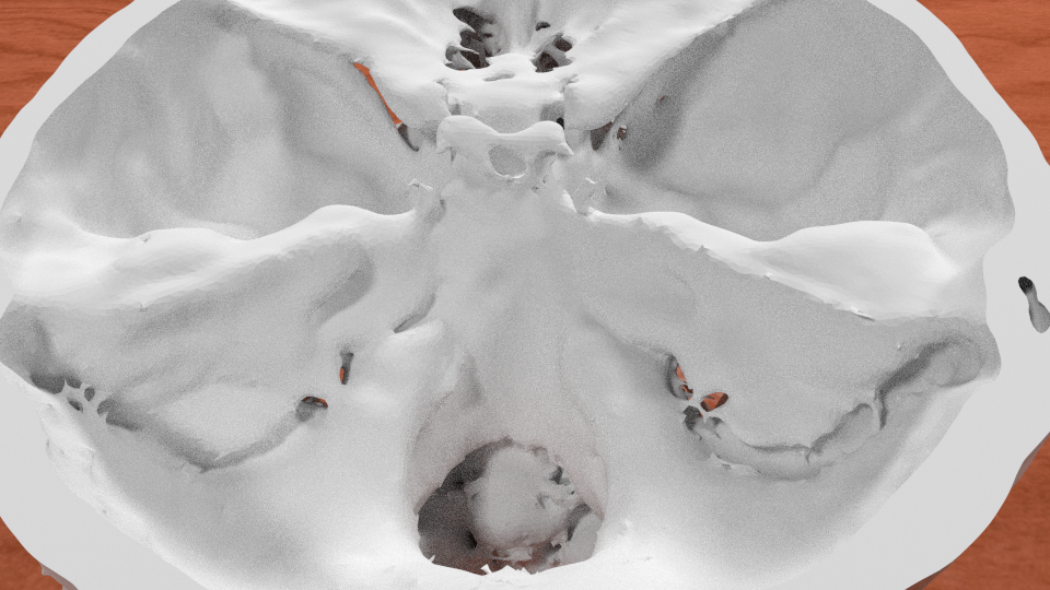
 The base of the skull is one of the most complex and difficult parts of the body for doctors in training to master. And one of the most important. It is comprised of multiple bones (the ethmoid, sphenoid, occipital, frontal, parietal, and temporal, to be exact) and has numerous foramina (holes) through which arteries, veins, and the vital cranial nerves and spinal cord exit the skull on their way to and from the body.
These structures, although very small, are critically important clinically. Compromise of a tiny foramen (hole) can lead to deafness, blindness, paralysis, or even stroke or death. Because of the importance of this small space, medical students around the world struggle to learn the complexities and subtleties of skull base anatomy.
Unfortunately, pictures in an anatomy book just don't cut it. Real human skulls can demonstrate this anatomy well, but these are expensive and the skull has to be cut and opened in order to display the relevant anatomy. This is why I created a 3D printed skull base from real CT scan data.
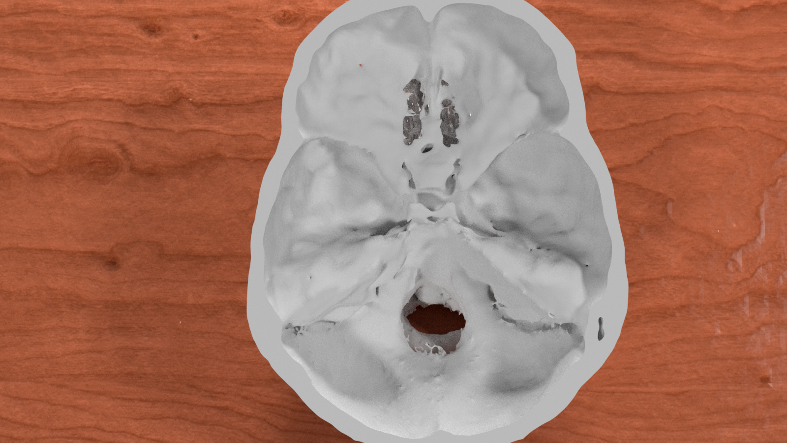
Available in full size and half-size models, the skull base exhibits exquisite anatomical detail. Digital files of the skull base are available for free download in full size (STL, COLLADA) and half-size (STL, COLLADA) versions.
Very high resolution prints are available for a fee at Shapeways in both full-size and half-size. The half-size model is quite inexpensive so you don't have to worry if it is damaged by rough handling of multiple students.
In the near future I will be posting more anatomical digital models.
For updates on news and new blog entries, follow us on Twitter at @Embodi3D


5 Comments
Recommended Comments
Create an account or sign in to comment
You need to be a member in order to leave a comment
Create an account
Sign up for a new account in our community. It's easy!
Register a new accountSign in
Already have an account? Sign in here.
Sign In Now