
Search the Community
Showing results for tags 'socket and ball joint'.
Found 24 results
-

Version 1.0.0
17 downloads
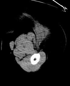
The bony pelvis is formed by 4 bones; a pair of hip bones, the sacrum and the coccyx. The bony pelvis supports the pelvic viscera and works to transmit force from the axial skeleton to the lower limbs. The two hip bones are related anteriorly by the symphysis pubis and posteriorly to the sacroiliac joints bilaterally. The hip joint is a large synovial socket and ball joint which is formed by the femoral head (the ball) and the acetabulum (the socket). The acetabulum is formed by pelvic bones; the ilium, the ischium and the pubis. The hip joint represents the articulation between the lower extremity and the axial skeleton and allows a high degree of mobility while being stable. The CT scan is derived from the file STS_040 The 3D bone model created from this scan can be reviewed at: The 3D muscle model created from this scan can be reviewed at: The 3D skin model created from this scan can be reviewed at:Free -


Version 1.0.0
32 downloads
The bony pelvis is formed by 4 bones; a pair of hip bones, the sacrum and the coccyx. The bony pelvis supports the pelvic viscera and works to transmit force from the axial skeleton to the lower limbs. The two hip bones are related anteriorly by the symphysis pubis and posteriorly to the sacroiliac joints bilaterally. The hip joint is a large synovial socket and ball joint which is formed by the femoral head (the ball) and the acetabulum (the socket). The acetabulum is formed by pelvic bones; the ilium, the ischium and the pubis. The hip joint represents the articulation between the lower extremity and the axial skeleton and allows a high degree of mobility while being stable. This 3D model was created from the file STS_040 The original CT examination can be reviewed at: The 3D muscle model created from this scan can be reviewed at: The 3D skin model created from this scan can be reviewed at:Free -

Version 1.0.0
37 downloads
The bony pelvis is formed by 4 bones; a pair of hip bones, the sacrum and the coccyx. The bony pelvis supports the pelvic viscera and works to transmit force from the axial skeleton to the lower limbs. The two hip bones are related anteriorly by the symphysis pubis and posteriorly to the sacroiliac joints bilaterally. The hip joint is a large synovial socket and ball joint which is formed by the femoral head (the ball) and the acetabulum (the socket). The acetabulum is formed by pelvic bones; the ilium, the ischium and the pubis. The hip joint represents the articulation between the lower extremity and the axial skeleton and allows a high degree of mobility while being stable. This 3D model was created from the file STS_040 The original CT examination can be reviewed at: The 3D bone model created from this scan can be reviewed at: The 3D skin model created from this scan can be reviewed at: This model shows a case of epithelioid sarcoma, which can be viewed at:Free -

Version 1.0.0
20 downloads
The bony pelvis is formed by 4 bones; a pair of hip bones, the sacrum and the coccyx. The bony pelvis supports the pelvic viscera and works to transmit force from the axial skeleton to the lower limbs. The two hip bones are related anteriorly by the symphysis pubis and posteriorly to the sacroiliac joints bilaterally. The hip joint is a large synovial socket and ball joint which is formed by the femoral head (the ball) and the acetabulum (the socket). The acetabulum is formed by pelvic bones; the ilium, the ischium and the pubis. The hip joint represents the articulation between the lower extremity and the axial skeleton and allows a high degree of mobility while being stable. This 3D model was created from the file STS_040 The original CT examination can be reviewed at: The 3D bone model created from this scan can be reviewed at: The 3D muscle model created from this scan can be reviewed at:Free -

Version 1.0.0
5 downloads
The hip joint is a large synovial socket and ball joint which is formed by the femoral head (the ball) and the acetabulum (the socket). The acetabulum is formed by pelvic bones; the ilium, the ischium and the pubis. The hip joint represents the articulation between the lower extremity and the axial skeleton and allows a high degree of mobility while being stable. The CT scan is derived from the file STS_040 The 3D bone model created from this scan can be reviewed at: The 3D muscle model created from this scan can be reviewed at:Free -

Version 1.0.0
7 downloads
The hip joint is a large synovial socket and ball joint which is formed by the femoral head (the ball) and the acetabulum (the socket). The acetabulum is formed by pelvic bones; the ilium, the ischium and the pubis. The hip joint represents the articulation between the lower extremity and the axial skeleton and allows a high degree of mobility while being stable. This 3D model was created from the file STS_040 The original CT examination can be reviewed at: The 3D muscle model created from this scan can be reviewed at:Free -

Version 1.0.0
8 downloads
The hip joint is a large synovial socket and ball joint which is formed by the femoral head (the ball) and the acetabulum (the socket). The acetabulum is formed by pelvic bones; the ilium, the ischium and the pubis. The hip joint represents the articulation between the lower extremity and the axial skeleton and allows a high degree of mobility while being stable. The muscles of the hip consist of four main groups Gluteal group: the gluteus maximus, gluteus medius, gluteus minimus and tensor fasciae latae Adductor group: the adductor brevis, adductor longus, adductor magnus, pectineus and gracilis Iliopsoas group: the iliacus and psoas major Lateral rotator group: the externus and internus obturators, the piriformis, the superior and inferior gemelli and the quadratus femoris Other hip muscles: the rectus femoris and the sartorius This 3D model was created from the file STS_040 The original CT examination can be reviewed at: The 3D bone model created from this scan can be reviewed at:Free -
Version 1.0.0
5 downloads
The hip joint is a large synovial socket and ball joint which is formed by the femoral head (the ball) and the acetabulum (the socket). The acetabulum is formed by pelvic bones; the ilium, the ischium and the pubis. The hip joint represents the articulation between the lower extremity and the axial skeleton and allows a high degree of mobility while being stable. The CT scan is derived from the file STS_040 The 3D bone model created from this scan can be reviewed at: The 3D muscle model created from this scan can be reviewed at:Free -

Version 1.0.0
6 downloads
The hip joint is a large synovial socket and ball joint which is formed by the femoral head (the ball) and the acetabulum (the socket). The acetabulum is formed by pelvic bones; the ilium, the ischium and the pubis. The hip joint represents the articulation between the lower extremity and the axial skeleton and allows a high degree of mobility while being stable. This 3D model was created from the file STS_040 The original CT examination can be reviewed at: The 3D muscle model created from this scan (showing a case of epithelioid sarcoma with vascular differentiation) can be reviewed at:Free -

Version 1.0.0
1 download
The bony pelvis is formed by 4 bones; a pair of hip bones, the sacrum and the coccyx. The bony pelvis supports the pelvic viscera and works to transmit force from the axial skeleton to the lower limbs. The two hip bones are related anteriorly by the symphysis pubis and posteriorly to the sacroiliac joints bilaterally. The hip joint is a large synovial socket and ball joint which is formed by the femoral head (the ball) and the acetabulum (the socket). The acetabulum is formed by pelvic bones; the ilium, the ischium and the pubis. The hip joint represents the articulation between the lower extremity and the axial skeleton and allows a high degree of mobility while being stable. This model shows parts of the fingers as the patient's hands were set beside the body. The CT scan is derived from the file STS_037 The original CT examination can be reviewed at: The 3D bone model created from this scan can be reviewed at: The 3D muscle model created from this scan can be reviewed at:Free -

Version 1.0.0
14 downloads
The bony pelvis is formed by 4 bones; a pair of hip bones, the sacrum and the coccyx. The bony pelvis supports the pelvic viscera and works to transmit force from the axial skeleton to the lower limbs. The two hip bones are related anteriorly by the symphysis pubis and posteriorly to the sacroiliac joints bilaterally. The hip joint is a large synovial socket and ball joint which is formed by the femoral head (the ball) and the acetabulum (the socket). The acetabulum is formed by pelvic bones; the ilium, the ischium and the pubis. The hip joint represents the articulation between the lower extremity and the axial skeleton and allows a high degree of mobility while being stable. This model shows parts of the fingers as the patient's hands were set beside the body. The CT scan is derived from the file STS_037 The original CT examination can be reviewed at: The 3D muscle model created from this scan can be reviewed at: The 3D skin model created from this scan can be reviewed at:Free -

Version 1.0.0
22 downloads
The bony pelvis is formed by 4 bones; a pair of hip bones, the sacrum and the coccyx. The bony pelvis supports the pelvic viscera and works to transmit force from the axial skeleton to the lower limbs. The two hip bones are related anteriorly by the symphysis pubis and posteriorly to the sacroiliac joints bilaterally. The hip joint is a large synovial socket and ball joint which is formed by the femoral head (the ball) and the acetabulum (the socket). The acetabulum is formed by pelvic bones; the ilium, the ischium and the pubis. The hip joint represents the articulation between the lower extremity and the axial skeleton and allows a high degree of mobility while being stable. The CT scan is derived from the file STS_037 The 3D bone model created from this scan can be reviewed at: The 3D muscle model created from this scan can be reviewed at: The 3D skin model created from this scan can be reviewed at:Free -

Version 1.0.0
8 downloads
The bony pelvis is formed by 4 bones; a pair of hip bones, the sacrum and the coccyx. The bony pelvis supports the pelvic viscera and works to transmit force from the axial skeleton to the lower limbs. The two hip bones are related anteriorly by the symphysis pubis and posteriorly to the sacroiliac joints bilaterally. The hip joint is a large synovial socket and ball joint which is formed by the femoral head (the ball) and the acetabulum (the socket). The acetabulum is formed by pelvic bones; the ilium, the ischium and the pubis. The hip joint represents the articulation between the lower extremity and the axial skeleton and allows a high degree of mobility while being stable. The muscles of the hip consist of four main groups; Gluteal group: the gluteus maximus, gluteus medius, gluteus minimus and tensor fasciae latae Adductor group: the adductor brevis, adductor longus, adductor magnus, pectineus and gracilis Iliopsoas group: the iliacus and psoas major Lateral rotator group: the externus and internus obturators, the piriformis, the superior and inferior gemelli and the quadratus femoris Other hip muscles: the rectus femoris and the sartorius The CT scan is derived from the file STS_037 The original CT examination can be reviewed at: The 3D bone model created from this scan can be reviewed at: The 3D skin model created from this scan can be reviewed at:Free -

Version 1.0.0
3 downloads
The hip joint is a large synovial socket and ball joint which is formed by the femoral head (the ball) and the acetabulum (the socket). The acetabulum is formed by pelvic bones; the ilium, the ischium and the pubis. The hip joint represents the articulation between the lower extremity and the axial skeleton and allows a high degree of mobility while being stable. The CT scan is derived from the file STS_037 The 3D bone model created from this scan can be reviewed at: The 3D muscle model created from this scan can be reviewed at:Free -

Version 1.0.0
7 downloads
The hip joint is a large synovial socket and ball joint which is formed by the femoral head (the ball) and the acetabulum (the socket). The acetabulum is formed by pelvic bones; the ilium, the ischium and the pubis. The hip joint represents the articulation between the lower extremity and the axial skeleton and allows a high degree of mobility while being stable. This model shows parts of the fingers as the patient's hand was set beside the body. This 3D model was created from the file STS_037 The original CT examination can be reviewed at: The 3D muscle model created from this scan can be reviewed at:Free -

Version 1.0.0
3 downloads
The hip joint is a large synovial socket and ball joint which is formed by the femoral head (the ball) and the acetabulum (the socket). The acetabulum is formed by pelvic bones; the ilium, the ischium and the pubis. The hip joint represents the articulation between the lower extremity and the axial skeleton and allows a high degree of mobility while being stable. The muscles of the hip consist of four main groups; Gluteal group: the gluteus maximus, gluteus medius, gluteus minimus and tensor fasciae latae Adductor group: the adductor brevis, adductor longus, adductor magnus, pectineus and gracilis Iliopsoas group: the iliacus and psoas major Lateral rotator group: the externus and internus obturators, the piriformis, the superior and inferior gemelli and the quadratus femoris Other hip muscles: the rectus femoris and the sartorius This model shows parts of the fingers as the patient's hand was set beside the body. This 3D model was created from the file STS_037 The original CT examination can be reviewed at: The 3D bone model created from this scan can be reviewed at:Free -

Version 1.0.0
5 downloads
The hip joint is a large synovial socket and ball joint which is formed by the femoral head (the ball) and the acetabulum (the socket). The acetabulum is formed by pelvic bones; the ilium, the ischium and the pubis. The hip joint represents the articulation between the lower extremity and the axial skeleton and allows a high degree of mobility while being stable. The CT scan is derived from the file STS_037 The 3D bone model created from this scan can be reviewed at: The 3D muscle model created from this scan can be reviewed at:Free -

Version 1.0.0
1 download
The hip joint is a large synovial socket and ball joint which is formed by the femoral head (the ball) and the acetabulum (the socket). The acetabulum is formed by pelvic bones; the ilium, the ischium and the pubis. The hip joint represents the articulation between the lower extremity and the axial skeleton and allows a high degree of mobility while being stable. This model shows parts of the fingers as the patient's hand was set beside the body. This 3D model was created from the file STS_037 The original CT examination can be reviewed at: The 3D muscle model created from this scan can be reviewed at:Free -

Version 1.0.0
0 downloads
The hip joint is a large synovial socket and ball joint which is formed by the femoral head (the ball) and the acetabulum (the socket). The acetabulum is formed by pelvic bones; the ilium, the ischium and the pubis. The hip joint represents the articulation between the lower extremity and the axial skeleton and allows a high degree of mobility while being stable. This CT scan was cropped from the file STS_036 The 3D bone model created from this scan can be reviewed at: The 3D muscle model created from this scan can be reviewed at:Free -

Version 1.0.0
6 downloads
The hip joint is a large synovial socket and ball joint which is formed by the femoral head (the ball) and the acetabulum (the socket). The acetabulum is formed by pelvic bones; the ilium, the ischium and the pubis. The hip joint represents the articulation between the lower extremity and the axial skeleton and allows a high degree of mobility while being stable. This 3D model was created from the file STS_036 The original CT examination can be reviewed at: The 3D muscle model created from this scan can be reviewed at:Free -

Version 1.0.0
4 downloads
The hip joint is a large synovial socket and ball joint which is formed by the femoral head (the ball) and the acetabulum (the socket). The acetabulum is formed by pelvic bones; the ilium, the ischium and the pubis. The hip joint represents the articulation between the lower extremity and the axial skeleton and allows a high degree of mobility while being stable. The muscles of the hip consist of four main groups Gluteal group: the gluteus maximus, gluteus medius, gluteus minimus, and tensor fasciae latae Adductor group: the adductor brevis, adductor longus, adductor magnus, pectineus, and gracilis Iliopsoas group: the iliacus and psoas major Lateral rotator group: the externus and internus obturators, the piriformis, the superior and inferior gemelli, and the quadratus femoris Other hip muscles: the rectus femoris and the sartorius This 3D model was created from the file STS_036 The original CT examination can be reviewed at: The 3D bone model created from this scan can be reviewed at:Free -

Version 1.0.0
2 downloads
The hip joint is a large synovial socket and ball joint which is formed by the femoral head (the ball) and the acetabulum (the socket). The acetabulum is formed by pelvic bones; the ilium, the ischium and the pubis. The hip joint represents the articulation between the lower extremity and the axial skeleton and allows a high degree of mobility while being stable. This 3D model was created from the file STS_036 The original CT examination can be reviewed at: The 3D muscle model created from this scan can be reviewed at:Free -

Version 1.0.0
1 download
The hip joint is a large synovial socket and ball joint which is formed by the femoral head (the ball) and the acetabulum (the socket). The acetabulum is formed by pelvic bones; the ilium, the ischium and the pubis. The hip joint represents the articulation between the lower extremity and the axial skeleton and allows a high degree of mobility while being stable. This CT scan was cropped from the file STS_036 The 3D bone model created from this scan can be reviewed at: The 3D muscle model created from this scan can be reviewed at:Free


