
Search the Community
Showing results for tags 'column'.
-

Version 1.0.0
11 downloads
This kidney model is derived from a hgih resolution CT scan of the abdomen and captures the renal cortex, renal columns, aorta and renal arteries. The model consists of a single STL file with ~500k triangles.$2.99 -

Version 1.0.0
12 downloads
T12 FRACTURE WITRH CANAL INVASION, NEUROLOGICAL ASIA C, abdomen, pelvis, .stl, 3d, model, printing, medical, medicine, cysts, spleen, liver, pancreas, kidney, small, bowel, colon, spinous, transverse, spine, lumbar, pelvis, iliac, pubis, foramen, sacrum, foramina, fracture, column, femur, neck, head, hip, ct, axial, dicom, without, contrast,Free -

Version 1.0.0
7 downloads
T12 FRACTURE WITRH CANAL INVASION, NEUROLOGICAL ASIA C, column, fracture, abdomen, ct, scan, without, contrast, .stl, 3d, model, printing, medical, medicine, diaphragm, lumbar, spine, ribs, kidney, small, bowel, colon, transverse, cyst, spleen, liver, T12, bone, muscle, iliac, pelvis, sacroiliac, joint, foramen, foramina, coccyx, sigmoid, pancreas, tail, urinary, bladder,Free -
0 downloads
Extracted from CT., hip and spine, lumbar, spine, columb, vertebrae, stl, bone, print, 3d model hip, and, spine, acetabulum, pelvis, iliac, ribs, thorax, bone, 3d, model, .stl, printable, intervertebral, disc, body, transverse, spinous, foramen, laminae, facet, joint, costovertebral, chest,Free -
-

Version 1.0.0
36 downloads
cat scan - stl file processed, back, leg, pelvis, stl, 3dmodel, bone, vetrinary, tail, column, lumbarFree -

Version 1.0.0
1 download
Healthy head Lateral ventricle (frontal horn), Circular sulcus of insula, Head of caudate nucleus, Lateral sulcus, Internal capsule (anterior limb), Precentral gyrus, Cave of septum pellucidum, Central sulcus, Globus pallidus, Postcentral gyrus, Insula, Cisterns, Fornix (column), Interventricular foramen (foramen of Monro), Third ventricle, Claustrum, Thalamus, Putamen, Superior temporal gyrus, Extreme capsule, Internal cerebral vein, External capsule Tail of caudate nucleus, Choroid plexus in trigone of posterior horn of lateral ventricle, great cerebral vein, Corpus callosum, splenium, Straight sinus Middle temporal gyrus, Parieto-occipital sulcus, Parietal bone, Occipital gyri, Cuneus, Striate cortex, cerebellumFree -

Version 1.0.0
1 download
Core1 - stl file processed This file was created with democratiz3D. Automatically create 3D printable models from CT scans. Learn more. column, vertebrae, 3d, model, stl, bone, lumbar, spine, body, printableFree -

Version 1.0.0
11 downloads
This 3D printable STL file contains a model of the lumbar spine was derived from a medical CT scan. It shows a compression fracture at L5. This model was created using the democratiz3D 3D model creation service 0522c0878 L5fxFree -
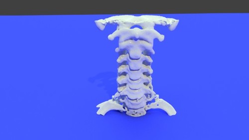
Version 1.0.0
65 downloads
This 3D printable STL file contains a model of the cervical spine was derived from a medical CT scan and shows the spine in great detail. This model was created using the democratiz3D 3D model creation service 0522c0883Free -

Version 1.0.0
8 downloads
This 3D printable STL file contains a model of the thoracic spine derived from a CT. This model was created using the democratiz3D 3D model creation service TCGA-CS-6185Free -

Version 1.0.0
88 downloads
This 3D printable STL file contains a model of the thoracic spine derived from a CT. The spine has significant scoliosis (abnormal curvature) This model was created using the democratiz3D 3D model creation service TCGA-DD-A1E9 thorax with scoliosis - processedFree -

28 downloads
This model is a cervical spine of a dog who has a tumor invading the lower cervical spine. Model details File format: STL Vertices 423273 Triangles 846734 Mesh is manifold (watertight and error-free) and ready for 3D printing. This model was created using the democratiz3D service.Free -

Version 1.0.0
1 download
A CT of a small section of the normal lumbar spine from a dog, k9, lumbar, column, bone, canine, vertebrae, animal, axial, dicomFree -

Version 1.0.0
0 downloads
Canine Lumbar Vertebrae - stl file processed, lumbar, stl, spine, column, vertebrae, stl, 3dmodel, k9, veterinary, canineFree -

Version 1.0.0
1 download
J Rodriguez Thoracic 10 2.5 ct with contrast, chest, dorsal, spine, column, bone, 3dmodels, stl, axialFree -

Version 1.0.0
9 downloads
9 chest c 7 - stl file processed bone, 3dmodel, stl, spine, column, chest, thorax, ribs, clavicleFree -


Version 1.0.0
38 downloads
This whole body bone STL file ready for medical 3D printing including chest, abdomen and pelvis was converted from an NRRD file to an STL file using democratiz3D, embodi3D's file conversion service. The chest wall (thoracic cage) is composed by twelve pairs of ribs laterally and the sternum anteriorly. The ribs are attached to the dorsal vertebrae (thoracic spine) posteriorly and along their costal cartilage to the sternum. The thoracic cage main function is to protect the vital chest organs such as the heart and lungs. The cervical spine is the upper most spines forming the spinal column, extending from the skull base to the level of the thoracic vertebra (the spines with attached ribs). The cervical spines are usually seven and the main function is to support the skull and to protect the spinal cord. The dorsal (thoracic) spine forms the middle portion of the vertebral column extending below the seventh cervical vertebra to above the first lumbar vertebra. The dorsal spine is formed by twelve vertebral bodies. The vertebrae forming the dorsal spine are unique in shape as they are the only vertebral bodies articulating with ribs. The lumbar spine represents the mid-lower segment of the vertebral column and is composed of five adjacent vertebrae. They are convex anteriorly to form a lumbar lordosis. The lumbar spine facet joints allows limited movements and rotation. The bony pelvis is formed by 4 bones; a pair of hip bones, the sacrum and the coccyx. The bony pelvis supports the pelvic viscera and works to transmit force from the axial skeleton to the lower limbs. The two hip bones are related anteriorly by the symphysis pubis and posteriorly to the sacroiliac joints bilaterally. This 3D model was created from the file ABD_LYMPH_001 The original CT examination can be reviewed at: The 3D muscle model created from this scan can be reviewed at:Free -

Version 1.0.0
224 downloads
Whole body NRRD file converted from CT Scan for Medical 3D Printing includes the chest, abdomen and pelvis. The chest wall (thoracic cage) is composed by twelve pairs of ribs laterally and the sternum anteriorly. The ribs are attached to the dorsal vertebrae (thoracic spine) posteriorly and along their costal cartilage to the sternum. The thoracic cage main function is to protect the vital chest organs such as the heart and lungs. The cervical spine is the upper most spines forming the spinal column, extending from the skull base to the level of the thoracic vertebra (the spines with attached ribs). The cervical spines are usually seven and the main function is to support the skull and to protect the spinal cord. The dorsal (thoracic) spine forms the middle portion of the vertebral column extending below the seventh cervical vertebra to above the first lumbar vertebra. The dorsal spine is formed by twelve vertebral bodies. The vertebrae forming the dorsal spine are unique in shape as they are the only vertebral bodies articulating with ribs. The lumbar spine represents the mid-lower segment of the vertebral column and is composed of five adjacent vertebrae. They are convex anteriorly to form a lumbar lordosis. The lumbar spine facet joints allows limited movements and rotation. The bony pelvis is formed by 4 bones; a pair of hip bones, the sacrum and the coccyx. The bony pelvis supports the pelvic viscera and works to transmit force from the axial skeleton to the lower limbs. The two hip bones are related anteriorly by the symphysis pubis and posteriorly to the sacroiliac joints bilaterally. The CT scan is derived from the file ABD_LYMPH_001 The 3D bone model created from this scan can be reviewed at: The 3D muscle model created from this scan can be reviewed at:Free -

Version 1.0.0
10 downloads
The dorsal (thoracic) spine forms the middle portion of the vertebral column extending below the seventh cervical vertebra to above the first lumbar vertebra. The dorsal spine is formed by twelve vertebral bodies. The vertebrae forming the dorsal spine are unique in shape as they are the only vertebral bodies articulating with ribs. This 3D model was created from the file ABD_LYMPH_001 The original CT examination can be reviewed at:Free -


Version 1.0.0
1 download
The chest wall (thoracic cage) is composed by twelve pairs of ribs laterally and the sternum anteriorly. The ribs are attached to the dorsal vertebrae (thoracic spine) posteriorly and along their costal cartilage to the sternum. The thoracic cage main function is to protect the vital chest organs such as the heart and lungs. There are five muscles that make up the thoracic cage; the intercostals (external, internal and innermost), subcostals, and transversus thoracis. This 3D model was created from the file ABD_LYMPH_002 The original CT examination can be reviewed at: The 3D bone model created from this scan can be reviewed at: The 3D skin model created from this scan can be reviewed at:Free -

Version 1.0.0
1 download
The chest wall (thoracic cage) is composed by twelve pairs of ribs laterally and the sternum anteriorly. The ribs are attached to the dorsal vertebrae (thoracic spine) posteriorly and along their costal cartilage to the sternum. The thoracic cage main function is to protect the vital chest organs such as the heart and lungs. There are five muscles that make up the thoracic cage; the intercostals (external, internal and innermost), subcostals, and transversus thoracis. This 3D model was created from the file ABD_LYMPH_002 The original CT examination can be reviewed at: The 3D bone model created from this scan can be reviewed at: The 3D muscle model created from this scan can be reviewed at:Free -

Version 1.0.0
0 downloads
The chest wall (thoracic cage) is composed by twelve pairs of ribs laterally and the sternum anteriorly. The ribs are attached to the dorsal vertebrae (thoracic spine) posteriorly and along their costal cartilage to the sternum. The thoracic cage main function is to protect the vital chest organs such as the heart and lungs. There are five muscles that make up the thoracic cage; the intercostals (external, internal and innermost), subcostals, and transversus thoracis. The CT scan is derived from the file ABD_LYMPH_002 The 3D bone model created from this scan can be reviewed at: The 3D muscle model created from this scan can be reviewed at: The 3D skin model created from this scan can be reviewed at:Free -

Version 1.0.0
0 downloads
The lumbar spine represents the mid-lower segment of the vertebral column and is composed of five adjacent vertebrae. They are convex anteriorly to form a lumbar lordosis. The lumbar spine facet joints allows limited movements and rotation. Each lumbar vertebra is formed of: A body which is kidney shaped and is convex anteriorly while flattened posteriorly, pedicles and lamina, transverse processes, articular processes and a spinous process. This model shows the whole vertebral body with related articulations of the lower level vertebra The CT scan is derived from the file ABD_LYMPH_002 The 3D bone model created from this scan can be reviewed at:Free -

Version 1.0.0
0 downloads
The lumbar spine represents the mid-lower segment of the vertebral column and is composed of five adjacent vertebrae. They are convex anteriorly to form a lumbar lordosis. The lumbar spine facet joints allows limited movements and rotation. Each lumbar vertebra is formed of: A body which is kidney shaped and is convex anteriorly while flattened posteriorly, pedicles and lamina, transverse processes, articular processes and a spinous process. This model shows the whole vertebral body with related articulations of the lower level vertebra as well as the contrast media within the related vasculature and bowels. This 3D model was created from the file ABD_LYMPH_002 The original CT examination can be reviewed at:Free


