


embodi3d
-
Posts
840 -
Joined
-
Last visited
-
Days Won
41
Content Type
Profiles
Blogs
Forums
Downloads
Gallery
Store
Events
Files posted by embodi3d
-

Free
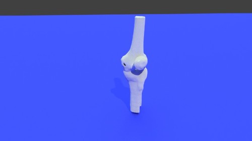
Right knee - Skin model STL file from converted CT scan
The knee joint is formed by three bones: the femur, the tibia and the patella. the knee joint is the largest synovial joint and provides the flexion and extension movements of the leg as well as relative medial and lateral rotations while in relative flexion.
The knee joint articulations are two condylar joints between the femur and the tibia as well as a joint between the patella and the femur. Although the fibula is closely related to the knee joint but it doesn't share in articulation. The knee joint is also formed by some ligaments and cartilage called (mensci) which are best imaged by MRI. This 3D model was created from the file STS_039 The original CT examination can be reviewed at: The 3D bone model created from this scan can be reviewed at: The 3D muscle model created from this scan can be reviewed at:17 downloads
- knee joint
- femur
- (and 10 more)
(0 reviews)0 comments
Updated
-
Free
Left knee - Bone model STL file from converted CT scan
By embodi3d in Extremity, Lower (Leg)
The knee joint is formed by three bones: the femur, the tibia and the patella. the knee joint is the largest synovial joint and provides the flexion and extension movements of the leg as well as relative medial and lateral rotations while in relative flexion.
The knee joint articulations are two condylar joints between the femur and the tibia as well as a joint between the patella and the femur. Although the fibula is closely related to the knee joint but it doesn't share in articulation. The knee joint is also formed by some ligaments and cartilage called (menisci) which are best imaged by MRI. This 3D model was created from the file STS_039 The original CT examination can be reviewed at: The 3D muscle model created from this scan can be reviewed at: The 3D skin model created from this scan can be reviewed at:12 downloads
- knee joint
- synovial joint
- (and 10 more)
(0 reviews)0 comments
Updated
-
Free
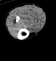
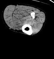
Left knee - CT scan
By embodi3d in Lower Extremity CTs
The knee joint is formed by three bones: the femur, the tibia and the patella. the knee joint is the largest synovial joint and provides the flexion and extension movements of the leg as well as relative medial and lateral rotations while in relative flexion.
The knee joint articulations are two condylar joints between the femur and the tibia as well as a joint between the patella and the femur. Although the fibula is closely related to the knee joint but it doesn't share in articulation. The knee joint is also formed by some ligaments and cartilage called (mensci) which are best imaged by MRI. The CT scan is derived from the file STS_039 The 3D bone model created from this scan can be reviewed at: The 3D muscle model created from this scan can be reviewed at: The 3D skin model created from this scan can be reviewed at:51 downloads
- knee joint
- femur
- (and 5 more)
(0 reviews)0 comments
Updated
-

Free
Right knee - Bone model STL file from converted CT scan
By embodi3d in Extremity, Lower (Leg)
The knee joint is formed by three bones: the femur, the tibia and the patella. the knee joint is the largest synovial joint and provides the flexion and extension movements of the leg as well as relative medial and lateral rotations while in relative flexion.
The knee joint articulations are two condylar joints between the femur and the tibia as well as a joint between the patella and the femur. Although the fibula is closely related to the knee joint but it doesn't share in articulation. The knee joint is also formed by some ligaments and cartilage called (mensci) which are best imaged by MRI. This 3D model was created from the file STS_039 The original CT examination can be reviewed at: The 3D muscle model created from this scan can be reviewed at: The 3D skin model created from this scan can be reviewed at:97 downloads
- knee joint
- synovial joint
- (and 10 more)
(0 reviews)0 comments
Updated
-
Free
Right knee - CT scan
By embodi3d in Lower Extremity CTs
The knee joint is formed by three bones: the femur, the tibia and the patella. the knee joint is the largest synovial joint and provides the flexion and extension movements of the leg as well as relative medial and lateral rotations while in relative flexion.
The knee joint articulations are two condylar joints between the femur and the tibia as well as a joint between the patella and the femur. Although the fibula is closely related to the knee joint but it doesn't share in articulation. The knee joint is also formed by some ligaments and cartilage called (mensci) which are best imaged by MRI. The CT scan is derived from the file STS_039 The 3D bone model created from this scan can be reviewed at: The 3D muscle model created from this scan can be reviewed at: The 3D skin model created from this scan can be reviewed at:277 downloads
- knee joint
- femur
- (and 5 more)
(0 reviews)0 comments
Updated
-

Free
Right foot - Skin model STL file from converted CT scan
The foot is a highly developed, biomechanically complex structure that serves to bear the weight of the body. The foot can be divided into 3 parts: the hindfoot, the midfoot, and the forefoot. The hindfoot is composed of 2 of the 7 tarsal bones, the talus, and the calcaneus; the midfoot contains the rest of the tarsal bones; and the forefoot contains the metatarsals and the phalanges. This 3D model was created from the file STS_039 The original CT examination can be reviewed at: The 3D bone model created from this scan can be reviewed at: The 3D muscle model created from this scan can be reviewed at:20 downloads
(0 reviews)0 comments
Updated
-

Free
Left foot - Muscle model STL file from converted CT scan
By embodi3d in Extremity, Lower (Leg) Muscles
The foot is a highly developed, biomechanically complex structure that serves to bear the weight of the body.
The foot can be divided into 3 parts: the hindfoot, the midfoot, and the forefoot. The hindfoot is composed of 2 of the 7 tarsal bones, the talus, and the calcaneus; the midfoot contains the rest of the tarsal bones; and the forefoot contains the metatarsals and the phalanges. This 3D model was created from the file STS_039 The original CT examination can be reviewed at: The 3D bone model created from this scan can be reviewed at: The 3D skin model created from this scan can be reviewed at:13 downloads
(0 reviews)0 comments
Updated
-

Free
Right foot - Muscle model STL file from converted CT scan
By embodi3d in Extremity, Lower (Leg) Muscles
The foot is a highly developed, biomechanically complex structure that serves to bear the weight of the body.
The foot can be divided into 3 parts: the hindfoot, the midfoot, and the forefoot. The hindfoot is composed of 2 of the 7 tarsal bones, the talus, and the calcaneus; the midfoot contains the rest of the tarsal bones; and the forefoot contains the metatarsals and the phalanges. This 3D model was created from the file STS_039 The original CT examination can be reviewed at: The 3D bone model created from this scan can be reviewed at: The 3D skin model created from this scan can be reviewed at:7 downloads
(0 reviews)0 comments
Updated
-

Free
Left foot - Bone model STL file from converted CT scan
By embodi3d in Extremity, Lower (Leg)
The foot is a highly developed, biomechanically complex structure that serves to bear the weight of the body.
The foot can be divided into 3 parts: the hindfoot, the midfoot, and the forefoot. The hindfoot is composed of 2 of the 7 tarsal bones, the talus, and the calcaneus; the midfoot contains the rest of the tarsal bones; and the forefoot contains the metatarsals and the phalanges. This 3D model was created from the file STS_039 The original CT examination can be reviewed at: The 3D muscle model created from this scan can be reviewed at: The 3D skin model created from this scan can be reviewed at:22 downloads
(0 reviews)0 comments
Updated
-

Free
Left foot - CT scan
By embodi3d in Lower Extremity CTs
The foot is a highly developed, biomechanically complex structure that serves to bear the weight of the body.
The foot can be divided into 3 parts: the hindfoot, the midfoot, and the forefoot. The hindfoot is composed of 2 of the 7 tarsal bones, the talus, and the calcaneus; the midfoot contains the rest of the tarsal bones; and the forefoot contains the metatarsals and the phalanges. The CT scan is derived from the file STS_039 The 3D bone model created from this scan can be reviewed at: The 3D muscle model created from this scan can be reviewed at: The 3D skin model created from this scan can be reviewed at:20 downloads
(0 reviews)0 comments
Updated
-

Free
Right foot - Bone model STL file from converted CT scan
By embodi3d in Extremity, Lower (Leg)
The foot is a highly developed, biomechanically complex structure that serves to bear the weight of the body.
The foot can be divided into 3 parts: the hindfoot, the midfoot, and the forefoot. The hindfoot is composed of 2 of the 7 tarsal bones, the talus, and the calcaneus; the midfoot contains the rest of the tarsal bones; and the forefoot contains the metatarsals and the phalanges. This 3D model was created from the file STS_039 The original CT examination can be reviewed at: The 3D muscle model created from this scan can be reviewed at: The 3D skin model created from this scan can be reviewed at:25 downloads
(0 reviews)0 comments
Updated
-

Free
Right foot - CT scan
By embodi3d in Lower Extremity CTs
The foot is a highly developed, biomechanically complex structure that serves to bear the weight of the body. The foot can be divided into 3 parts: the hindfoot, the midfoot, and the forefoot. The hindfoot is composed of 2 of the 7 tarsal bones, the talus, and the calcaneus; the midfoot contains the rest of the tarsal bones; and the forefoot contains the metatarsals and the phalanges. The CT scan is derived from the file STS_039 The 3D bone model created from this scan can be reviewed at: The 3D muscle model created from this scan can be reviewed at: The 3D skin model created from this scan can be reviewed at:13 downloads
(0 reviews)0 comments
Updated
-

Free
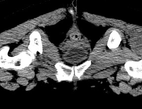
Pelvic Bones (female pelvis) - Muscle model STL file from converted CT scan
By embodi3d in Abdomen and Pelvis muscles
The bony pelvis is formed by 4 bones; a pair of hip bones, the sacrum and the coccyx. The bony pelvis supports the pelvic viscera and works to transmit force from the axial skeleton to the lower limbs.
The two hip bones are related anteriorly by the symphysis pubis and posteriorly to the sacroiliac joints bilaterally. This model if for a 57 years old female pelvis, it shows some irregular shaped pieces related to the contrast media within the colon as well as the upper halves of the femoral bones. The CT scan is derived from the file STS_040 The original CT examination can be reviewed at: The 3D bone model created from this scan can be reviewed at:16 downloads
(0 reviews)0 comments
Updated
-

Free
Chest wall - Muscle model STL file from converted CT scan
By embodi3d in Thorax and Ribs Muscles
The chest wall (thoracic cage) is composed by twelve pairs of ribs laterally and the sternum anteriorly. The ribs are attached to the dorsal vertebrae (thoracic spine) posteriorly and along their costal cartilage to the sternum. The thoracic cage main function is to protect the vital chest organs such as the heart and lungs. There are five muscles that make up the thoracic cage; the intercostals (external, internal and innermost), subcostals and transversus thoracis. This 3D model was created from the file STS_040 for a 57 years old female with breast implants. The original CT examination can be reviewed at: The 3D bone model created from this scan can be reviewed at: The 3D skin model created from this scan can be reviewed at:14 downloads
- chest wall
- thoracic cage
- (and 14 more)
(0 reviews)0 comments
Updated
-

Free
Chest wall - Skin model STL file from converted CT scan
The chest wall (thoracic cage) is composed by twelve pairs of ribs laterally and the sternum anteriorly. The ribs are attached to the dorsal vertebrae (thoracic spine) posteriorly and along their costal cartilage to the sternum.
The thoracic cage main function is to protect the vital chest organs such as the heart and lungs. There are five muscles that make up the thoracic cage; the intercostals (external, internal and innermost), subcostals and transversus thoracis. This 3D model was created from the file STS_040 for a 57 years old female with breast implants. The original CT examination can be reviewed at: The 3D bone model created from this scan can be reviewed at: The 3D muscle model created from this scan can be reviewed at:9 downloads
- chest wall
- thoracic cage
- (and 11 more)
(0 reviews)0 comments
Updated
-

Free
Pelvic Bones (female pelvis) - Bone model STL file from converted CT scan
By embodi3d in Spine and Pelvis
The bony pelvis is formed by 4 bones; a pair of hip bones, the sacrum and the coccyx. The bony pelvis supports the pelvic viscera and works to transmit force from the axial skeleton to the lower limbs.
The two hip bones are related anteriorly by the symphysis pubis and posteriorly to the sacroiliac joints bilaterally. This model if for a 57 years old female pelvis, it shows some irregular shaped pieces related to the contrast media within the colon as well as the upper halves of the femoral bones. The CT scan is derived from the file STS_040 The original CT examination can be reviewed at: The 3D muscle model created from this scan can be reviewed at:34 downloads
(0 reviews)0 comments
Updated
-
Free
Pelvic Bones (female pelvis) - CT Scan
By embodi3d in Abdomen and Pelvis CTs
The bony pelvis is formed by 4 bones; a pair of hip bones, the sacrum and the coccyx. The bony pelvis supports the pelvic viscera and works to transmit force from the axial skeleton to the lower limbs. The two hip bones are related anteriorly by the symphysis pubis and posteriorly to the sacroiliac joints bilaterally. This model if for a 57 years old female pelvis, it shows some irregular shaped pieces related to the contrast media within the colon as well as the upper halves of the femoral bones. The CT scan is derived from the file STS_040 The 3D bone model created from this scan can be reviewed at: The 3D muscle model created from this scan can be reviewed at:42 downloads
(0 reviews)0 comments
Updated
-

Free
Chest wall - Bone model STL file from converted CT scan
By embodi3d in Thorax and Ribs
The chest wall (thoracic cage) is composed by twelve pairs of ribs laterally and the sternum anteriorly. The ribs are attached to the dorsal vertebrae (thoracic spine) posteriorly and along their costal cartilage to the sternum. The thoracic cage main function is to protect the vital chest organs such as the heart and lungs. This 3D model was created from the file STS_040 The original CT examination can be reviewed at: The 3D muscle model created from this scan can be reviewed at: The 3D skin model created from this scan can be reviewed at:12 downloads
- chest wall
- thoracic cage
- (and 11 more)
(0 reviews)0 comments
Updated
-

Free
Chest wall - CT scan
By embodi3d in Thorax CTs
The chest wall (thoracic cage) is composed by twelve pairs of ribs laterally and the sternum anteriorly. The ribs are attached to the dorsal vertebrae (thoracic spine) posteriorly and along their costal cartilage to the sternum. The thoracic cage main function is to protect the vital chest organs such as the heart and lungs. The CT scan is derived from the file STS_040 The 3D bone model created from this scan can be reviewed at: The 3D muscle model created from this scan can be reviewed at: The 3D skin model created from this scan can be reviewed at:10 downloads
- chest wall
- thoracic cage
- (and 5 more)
(0 reviews)0 comments
Updated
-

Free
Right Shoulder - Muscle model STL file from converted CT scan
By embodi3d in Extremity, Upper (Arm) Muscles
The shoulder joint is a large and complex ball and socket joint formed by the humerus and the scapula (glenohumeral joint) while the clavicle join the acromion to form the acromioclavicular joint. The shoulder joint is the most mobile joint in the human body on cost of instability. Lot of elements share to compensate the instability such as rotator cuff muscles, tendons and ligaments as well as the glenoid labrum. Muscles of the shoulder joint
The rotator cuff: supraspinatus, infraspinatus, teres minor and subscapularis
Posterior muscle group: deltoid, latissimus dorsi and teres major
Anterior muscle group: pectoralis major and coracobrachialis This 3D model was created from the file STS_040 The original CT examination can be reviewed at: The 3D bone model created from this scan can be reviewed at:6 downloads
- shoulder joint
- ball and socket joint
- (and 21 more)
(0 reviews)0 comments
Updated
-

Free
Left Shoulder - Muscle model STL file from converted CT scan
By embodi3d in Extremity, Upper (Arm) Muscles
The shoulder joint is a large and complex ball and socket joint formed by the humerus and the scapula (glenohumeral joint) while the clavicle join the acromion to form the acromioclavicular joint.
The shoulder joint is the most mobile joint in the human body on cost of instability. Lot of elements share to compensate the instability such as rotator cuff muscles, tendons and ligaments as well as the glenoid labrum. Muscles of the shoulder joint
The rotator cuff: supraspinatus, infraspinatus, teres minor and subscapularis
Posterior muscle group: deltoid, latissimus dorsi and teres major
Anterior muscle group: pectoralis major and coracobrachialis This 3D model was created from the file STS_040 The original CT examination can be reviewed at: The 3D bone model created from this scan can be reviewed at:4 downloads
- shoulder joint
- ball and socket joint
- (and 21 more)
(0 reviews)0 comments
Updated
-

Free
Left Shoulder - Bone model STL file from converted CT scan
By embodi3d in Extremity, Upper (Arm)
The shoulder joint is a large and complex ball and socket joint formed by the humerus and the scapula (glenohumeral joint) while the clavicle join the acromion to form the acromioclavicular joint.
The shoulder joint is the most mobile joint in the human body on cost of instability. Lot of elements share to compensate the instability such as rotator cuff muscles, tendons and ligaments as well as the glenoid labrum. This 3D model was created from the file STS_040 The original CT examination can be reviewed at: The 3D muscle model created from this scan can be reviewed at:6 downloads
- shoulder joint
- ball and socket joint
- (and 12 more)
(0 reviews)0 comments
Updated
-

Free
Left Shoulder - CT scan
By embodi3d in Upper Extremity CTs
The shoulder joint is a large and complex ball and socket joint formed by the humerus and the scapula (glenohumeral joint) while the clavicle join the acromion to form the acromioclavicular joint. The shoulder joint is the most mobile joint in the human body on cost of instability. Lot of elements share to compensate the instability such as rotator cuff muscles, tendons and ligaments as well as the glenoid labrum. The CT scan is derived from the file STS_040 The 3D bone model created from this scan can be reviewed at: The 3D muscle model created from this scan can be reviewed at:10 downloads
- shoulder joint
- ball and socket joint
- (and 8 more)
(0 reviews)0 comments
Updated
-

Free
Right Shoulder - Bone model STL file from converted CT scan
By embodi3d in Extremity, Upper (Arm)
The shoulder joint is a large and complex ball and socket joint formed by the humerus and the scapula (glenohumeral joint) while the clavicle join the acromion to form the acromioclavicular joint. The shoulder joint is the most mobile joint in the human body on cost of instability. Lot of elements share to compensate the instability such as rotator cuff muscles, tendons and ligaments as well as the glenoid labrum. This 3D model was created from the file STS_040 The original CT examination can be reviewed at: The 3D muscle model created from this scan can be reviewed at:8 downloads
- shoulder joint
- ball and socket joint
- (and 12 more)
(0 reviews)0 comments
Updated
-

Free
Right Shoulder - CT scan
By embodi3d in Upper Extremity CTs
The shoulder joint is a large and complex ball and socket joint formed by the humerus and the scapula (glenohumeral joint) while the clavicle join the acromion to form the acromioclavicular joint. The shoulder joint is the most mobile joint in the human body on cost of instability. Lot of elements share to compensate the instability such as rotator cuff muscles, tendons and ligaments as well as the glenoid labrum. The CT scan is derived from the file STS_040 The 3D bone model created from this scan can be reviewed at: The 3D muscle model created from this scan can be reviewed at:9 downloads
- shoulder joint
- ball and socket joint
- (and 8 more)
(0 reviews)0 comments
Updated


