
-
Welcome to embodi3D Downloads! This is the largest and fastest growing library of 3D printable anatomic models generated from real medical scans on the Internet. A unique scientific resource, most of the material is free. Registered members can download, upload, and sell models. To convert your own medical scans to a 3D model, take a look at democratiz3D, our free and automated conversion service.
Alert (6/17/22) - The democratiz3D scan-to-model conversion app is down due to a technical issue. We are working on a solution.
Skin
Models of the skin and body surface
773 files
-

Free
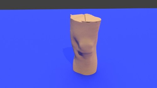
Right knee - Skin model STL file from converted CT scan
By embodi3d
The knee joint is formed by three bones: the femur, the tibia and the patella. the knee joint is the largest synovial joint and provides the flexion and extension movements of the leg as well as relative medial and lateral rotations while in relative flexion.
The knee joint articulations are two condylar joints between the femur and the tibia as well as a joint between the patella and the femur. Although the fibula is closely related to the knee joint but it doesn't share in articulation. The knee joint is also formed by some ligaments and cartilage called (menisci) which are best imaged by MRI. This 3D model was created from the file STS_051 The original CT examination can be reviewed at: The 3D bone model created from this scan can be reviewed at: The 3D muscle model created from this scan can be reviewed at:139 downloads
(0 reviews)0 comments
Updated
-

Free
Human hand - stl file processed
By JuanPolanco
Human hand - stl file processed
3dmodel, stl, finger, wrist, skin
123 downloads
(0 reviews)0 comments
Updated
-
.thumb.png.b400cec9de922ac238ad384b86876fc3.png)
Free
ear
By virat
please convert to stl file format , .stl, 3d, model, printable, ear, lobe, tragus, antitragus, skin
96 downloads
(0 reviews)0 comments
Updated
-

Free
Left foot - Skin model STL file from converted CT scan
By embodi3d
The foot is a highly developed, biomechanically complex structure that serves to bear the weight of the body.
The foot can be divided into 3 parts: the hindfoot, the midfoot, and the forefoot. The hindfoot is composed of 2 of the 7 tarsal bones, the talus, and the calcaneus; the midfoot contains the rest of the tarsal bones; and the forefoot contains the metatarsals and the phalanges. This 3D model was created from the file STS_039 The original CT examination can be reviewed at: The 3D bone model created from this scan can be reviewed at: The 3D muscle model created from this scan can be reviewed at:67 downloads
(1 review)0 comments
Updated
-

Free
Tutorial head and neck - processed
By Dr. Mike
3D printable head STL created from a CT scan. This model was created as part of this tutorial on created skin 3D printable models from medical CT scans.
62 downloads
(0 reviews)0 comments
Updated
-

Free
Hand - stl file processed
Hand - stl file processed
Have embodi3D 3D print this model for you. This file was created with democratiz3D. Automatically create 3D printable models from CT scans.
middle, finger, thumb, ring, pinky, index, wirst, palmar, dorsal, .stl, 3d, model, printable, skin, upper, limb
56 downloads
(0 reviews)0 comments
Updated
-
_nrrd.f9e5f4756f911424b16486964c4ff211_IIP9VK.stl_render1.thumb.jpg.e8eefffaae394db82c7b9acb4e88f388.jpg)
Free
Hand BST 3 skin - stl file processed
By ADM
Hand BST 3 skin - stl file processed
Have embodi3D 3D print this model for you. This file was created with democratiz3D. Automatically create 3D printable models from CT scans.
index, middle, ring little finger, thumb, skin, wrist, upper, limb, .stl, printable, 3d,
54 downloads
(0 reviews)0 comments
Updated
-

$5.50
(0 reviews)0 comments
Updated
-

Free
right-pelvis-2 - stl file processed
By nadi
right-pelvis-2 - stl file processed
3d model, stl, gluteus, pelvis, hip, vagina, genitalia, skin, tigh, femur, lumbar, spine, sacrum
This file was created with democratiz3D. Automatically create 3D printable models from CT scans. Learn more.
43 downloads
(0 reviews)0 comments
Updated
-

Free
Alexandra - stl file processed
By lew_is
Alexandra - stl file processed
Have embodi3D 3D print this model for you. This file was created with democratiz3D. Automatically create 3D printable models from CT scans.
upper, limb, wrist, palmar, dorsal, finger, index, thumb, anularis, pinky, medium, forearm, elbow, .stl, 3d, model, printable, printing, medical, medicine,
32 downloads
- upper limb
- wrist
- (and 17 more)
(0 reviews)0 comments
Updated
-

Free
foot and ankle Normal Left Foot and Ankle Skin Model 3D Printable STL File Converted from CT Scan
By embodi3d
This model is the left foot and ankle skin rendering of a 65-year-old male with left thigh myxoid fibrosarcoma. At the time of diagnosis, the patient had metastases to his lungs. The patient therefore underwent neoadjuvant radiotherapy, surgery, and adjuvant chemotherapy and was found to have an intermediate grade lesion at the time of diagnosis. The patient unfortunately died 9.5 months after diagnosis. This is an STL file created from DICOM images of his CT scan which may be used for 3D printing.
Topographical landmarks of the foot and ankle consist of muscular, tendinous, and bony structures. Proximally, the superficial muscles of the anterior (tibialis anterior), lateral (peroneals) and posterior (gastrocnemius) compartments may be palpated. Anteriorly, the tibialis anterior tendon crosses the ankle joint and is used as a landmark for ankle joint injections and aspirations, where the practitioner will place the needle just lateral to the tendon. Posteriorly, the gastrocnemius and soleus converge to form the Achilles tendon. Ruptures of the tendon as well as tendinous changes due to Achilles tendinopathy may be palpated. At the level of the ankle joint, the joint line, medial malleolus (distal tibia) and lateral malleolus (distal fibula) may be palpated. The extensor hallucis longus and extensor digitorum longus tendons are visible at the surface of the dorsal foot. The extensor digitorum brevis muscle belly is seen on the dorsum of the lateral foot. On the plantar foot, the plantar fascia may be palpated. Nodules associated with plantar fascial fibromatosis may be palpated here. Plantar fasciitis is also diagnosed when pain is associated with palpation of the insertion of the plantar fascia on the medial heel. Other common pathologies on the plantar foot are ulcerations associated with diabetic neuropathy and other neuropathic conditions.
This model was created from the file STS_023.
30 downloads
(0 reviews)0 comments
Updated
-
Free
Left knee - Skin model STL file from converted CT scan
By embodi3d
The knee joint is formed by three bones: the femur, the tibia and the patella. the knee joint is the largest synovial joint and provides the flexion and extension movements of the leg as well as relative medial and lateral rotations while in relative flexion.
The knee joint articulations are two condylar joints between the femur and the tibia as well as a joint between the patella and the femur. Although the fibula is closely related to the knee joint but it doesn't share in articulation. The knee joint is also formed by some ligaments and cartilage called (menisci) which are best imaged by MRI. This 3D model was created from the file STS_051 The original CT examination can be reviewed at: The 3D bone model created from this scan can be reviewed at: The 3D muscle model created from this scan can be reviewed at:28 downloads
(0 reviews)0 comments
Updated
-

Free
leg Right Leg Skin Model 3D Printable STL File Converted from CT Scan
By embodi3d
This model is the right leg skin rendering of a 65-year-old male with left thigh myxoid fibrosarcoma. At the time of diagnosis, the patient had metastases to his lungs. The patient therefore underwent neoadjuvant radiotherapy, surgery, and adjuvant chemotherapy and was found to have an intermediate grade lesion at the time of diagnosis. The patient is still living with the metastatic disease at 2.5 years since diagnosis. This is an STL file created from DICOM images of his CT scan which may be used for 3D printing.
Landmarks of the lower extremity consist of bony and muscular landmarks. Proximally, the extensor mechanism consists of the quadriceps tendon, patella, and the tibial tuberosity, which is located on the anterior proximal tibia, where the patellar tendon attaches. On the anteromedial surface of the tibia is Gerdy's tubercle, where the sartorius, gracilis, and semitendinosus attach. Laterally, the head of the fibula may be palpated, which is the attachment for the posterolateral corner structures of the knee joint. The peroneal nerve wraps around the fibular neck, and a tinel’s sign may be elicited due to its superficial position at this location.
Distally, the anterior ankle joint may be palpated. Pain with palpation may be indicative of osteoarthritis if general or an osteochondral defect if localized. The medial and lateral malleoli are located on either side of the tibiotalar joint, respectively and are the site of common ankle fractures. Posteriorly, the Achilles tendon inserts on the calcaneus. A defect along this tendon may be a sign of a tendon rupture. The superficial peroneal nerve can possibly be isolated on the lateral aspect of the dorsal foot with full plantarflexion of the fourth ray.
Topographical landmarks of the foot and ankle consist of muscular, tendinous, and bony structures. Proximally, the superficial muscles of the anterior (tibialis anterior), lateral (peroneals) and posterior (gastrocnemius) compartments may be palpated. Anteriorly, the tibialis anterior tendon crosses the ankle joint and is used as a landmark for ankle joint injections and aspirations, where the practitioner will place the needle just lateral to the tendon. Posteriorly, the gastrocnemius and soleus converge to form the Achilles tendon. Ruptures of the tendon as well as tendinous changes due to Achilles tendinopathy may be palpated. At the level of the ankle joint, the joint line, medial malleolus (distal tibia) and lateral malleolus (distal fibula) may be palpated. The extensor hallucis longus and extensor digitorum longus tendons are visible at the surface of the dorsal foot. The extensor digitorum brevis muscle belly is seen on the dorsum of the lateral foot. On the plantar foot, the plantar fascia may be palpated. Nodules associated with plantar fascial fibromatosis may be palpated here. Plantar fasciitis is also diagnosed when pain is associated with palpation of the insertion of the plantar fascia on the medial heel. Other common pathologies on the plantar foot are ulcerations associated with diabetic neuropathy and other neuropathic conditions.
This model was created from the file STS_022.
27 downloads
- lower extremity
- skin
- (and 8 more)
(0 reviews)0 comments
Updated
-

Free
Instructables 3D model of the leg skin - stl file processed
By MihaD
Instructables 3D model of the leg skin - stl file processed
Link to tutorial: https://www.instructables.com/How-to-Easily-and-Automatically-Convert-a-CT-Scan-/
Have embodi3D 3D print this model for you. This file was created with democratiz3D. Automatically create 3D printable models from CT scans.
lower, limb, .stl, 3d, model, printable, skin, thigh, gluteus, knee, ankle, pubis,
27 downloads
(0 reviews)0 comments
Updated
-

Free
foot and ankle Normal Left Foot and Ankle Skin Model 3D Printable STL File Converted from CT Scan
By embodi3d
This is the normal right foot and ankle skin model of a 56-year-old male with right anterior thigh pleomorphic leiomyosarcoma. This is an STL file created from DICOM images of his CT scan which may be used for 3D printing.
Topographical landmarks of the foot and ankle consist of muscular, tendinous, and bony structures. Proximally, the superficial muscles of the anterior (tibialis anterior), lateral (peroneals) and posterior (gastrocnemius) compartments may be palpated. Anteriorly, the tibialis anterior tendon crosses the ankle joint and is used as a landmark for ankle joint injections and aspirations, where the practitioner will place the needle just lateral to the tendon. Posteriorly, the gastrocnemius and soleus converge to form the Achilles tendon. Ruptures of the tendon, as well as tendinous changes due to Achilles tendinopathy, may be palpated. At the level of the ankle joint, the joint line, medial malleolus (distal tibia) and lateral malleolus (distal fibula) may be palpated. The extensor hallucis longus and extensor digitorum longus tendons are visible on the surface of the dorsal foot. The extensor digitorum brevis muscle belly is seen on the dorsum of the lateral foot. On the plantar foot, the plantar fascia may be palpated. Nodules associated with plantar fascial fibromatosis may be palpated here. Plantar fasciitis is also diagnosed when pain is associated with palpation of the insertion of the plantar fascia on the medial heel. Other common pathologies on the plantar foot are ulcerations associated with diabetic neuropathy and other neuropathic conditions.
This model was created from the file STS_014.
26 downloads
- lower extremity
- skin model
- (and 9 more)
(0 reviews)0 comments
Updated
-

Free
ea - stl file processed
By Zios
ea - stl file processed
Have embodi3D 3D print this model for you. This file was created with democratiz3D. Automatically create 3D printable models from CT scans.
Face, 3d, model, .stl, orbit, eye, nose, lips, head, skull, neck, temporal, frontal, parietal, maxilla, mandible, 3d, model, skin, ear, external auditory conduct,
25 downloads
(1 review)0 comments
Updated
-

Free
foot and ankle Normal Right Foot and Ankle Skin Model 3D Printable STL File Converted from CT Scan
By embodi3d
This model is the right foot and ankle skin rendering of a 65-year-old male with left thigh myxoid fibrosarcoma. At the time of diagnosis, the patient had metastases to his lungs. The patient therefore underwent neoadjuvant radiotherapy, surgery, and adjuvant chemotherapy and was found to have an intermediate grade lesion at the time of diagnosis. The patient unfortunately died 9.5 months after diagnosis. This is an STL file created from DICOM images of his CT scan which may be used for 3D printing.
Topographical landmarks of the foot and ankle consist of muscular, tendinous, and bony structures. Proximally, the superficial muscles of the anterior (tibialis anterior), lateral (peroneals) and posterior (gastrocnemius) compartments may be palpated. Anteriorly, the tibialis anterior tendon crosses the ankle joint and is used as a landmark for ankle joint injections and aspirations, where the practitioner will place the needle just lateral to the tendon. Posteriorly, the gastrocnemius and soleus converge to form the Achilles tendon. Ruptures of the tendon as well as tendinous changes due to Achilles tendinopathy may be palpated. At the level of the ankle joint, the joint line, medial malleolus (distal tibia) and lateral malleolus (distal fibula) may be palpated. The extensor hallucis longus and extensor digitorum longus tendons are visible at the surface of the dorsal foot. The extensor digitorum brevis muscle belly is seen on the dorsum of the lateral foot. On the plantar foot, the plantar fascia may be palpated. Nodules associated with plantar fascial fibromatosis may be palpated here. Plantar fasciitis is also diagnosed when pain is associated with palpation of the insertion of the plantar fascia on the medial heel. Other common pathologies on the plantar foot are ulcerations associated with diabetic neuropathy and other neuropathic conditions.
This model was created from the file STS_023.
25 downloads
(0 reviews)0 comments
Updated
-

Free
skin - stl file processed
skin - stl file processed
Have embodi3D 3D print this model for you. This file was created with democratiz3D. Automatically create 3D printable models from CT scans.
whole, body, skin, head, neck, thorax chest, abdomen, upper, lower, limbs, elbow, shoulder, back, thigh, knee, ankle, hip, pelvis, 3d, model, .stl, printable,
24 downloads
(0 reviews)0 comments
Updated
-

Free
Whole Body - Skin model STL file from converted CT scan
By embodi3d
Whole body: chest, abdomen and pelvis
The chest wall (thoracic cage) is composed by twelve pairs of ribs laterally and the sternum anteriorly. The ribs are attached to the dorsal vertebrae (thoracic spine) posteriorly and along their costal cartilage to the sternum.
The thoracic cage main function is to protect the vital chest organs such as the heart and lungs.
The cervical spine is the upper most spines forming the spinal column, extending from the skull base to the level of the thoracic vertebra (the spines with attached ribs). The cervical spines are usually seven and the main function is to support the skull and to protect the spinal cord.
The dorsal (thoracic) spine forms the middle portion of the vertebral column extending below the seventh cervical vertebra to above the first lumbar vertebra. The dorsal spine is formed by twelve vertebral bodies.
The vertebrae forming the dorsal spine are unique in shape as they are the only vertebral bodies articulating with ribs.
The lumbar spine represents the mid-lower segment of the vertebral column and is composed of five adjacent vertebrae. They are convex anteriorly to form a lumbar lordosis. The lumbar spine facet joints allows limited movements and rotation.
The bony pelvis is formed by 4 bones; a pair of hip bones, the sacrum and the coccyx. The bony pelvis supports the pelvic viscera and works to transmit force from the axial skeleton to the lower limbs.
The two hip bones are related anteriorly by the symphysis pubis and posteriorly to the sacroiliac joints bilaterally.
This 3D model was created from the file STS_040
The original CT examination can be reviewed at: The 3D bone model created from this scan can be reviewed at: The 3D muscle model created from this scan can be reviewed at:24 downloads
(0 reviews)0 comments
Updated
-
_nrrd.909eabac15c2d9eb90e59d91d7e34d31_NMBL0P.stl_render1.thumb.jpg.8e3e80d3fbc18af2f932332fa3f8b0c9.jpg)
Free
Nasopharynx Model (Skin) - stl file processed
Nasopharynx Model (Skin) - stl file processed
This file was created with democratiz3D. Automatically create 3D printable models from CT scans. Learn more.
3D, model, .stl, skin, .stl, orbit, eye, nose, lips, head, skull, neck, temporal, frontal, parietal, maxilla, mandible, 3d, model, skin, ear, external auditory conduct,
23 downloads
(0 reviews)0 comments
Updated
-

Free
Bane - stl file processed
By Pavle
Bane - stl file processed
Have embodi3D 3D print this model for you. This file was created with democratiz3D. Automatically create 3D printable models from CT scans.
upper, limb, wrist, palmar, dorsal, finger, index, thumb, anularis, pinky, medium, forearm, elbow, .stl, 3d, model, printable, printing, medical, medicine,
23 downloads
(0 reviews)0 comments
Updated
-

Free
Nemanja - stl file processed
By Pavle
Nemanja - stl file processed
Have embodi3D 3D print this model for you. This file was created with democratiz3D. Automatically create 3D printable models from CT scans.23 downloads
(0 reviews)0 comments
Submitted
-

$6.99
3D printable airway model from high resolution CT
By BioGuy
This model of the airway was segmented from a high resolution CT scan of a healthy young female and accurately captures the nasal passages, frontal sinuses, maxillary sinuses and ethmoid sinuses as well as the pharynx down to the epiglottis. The model includes two STL files. The first is the airway only and the second includes the exterior geometry of the nose and face with the airway Boolean subtracted from the interior:
The mesh is at a high resolution and consists of 600k triangles (airway only) and 1.3M triangles (face minus airway). The model is to scale in millimetres.
23 downloads
(0 reviews)0 comments
Updated
-

Free
Glitchy Face Skin - stl file processed
By glitchyg
Glitchy Face Skin - stl file processed
Have embodi3D 3D print this model for you. This file was created with democratiz3D. Automatically create 3D printable models from CT scans.
3d, model, .stl, printable, nasal, skin, zygomatic, arch, eyelid, lips, angle, ramus, body, ear, head, skull, neck, eye, surface, facial, maxillofacial,
22 downloads
(0 reviews)0 comments
Updated
-

Free
female phantom meditism - stl file processed
By EddeeNii
female phantom meditism - stl file processed, chest, female, breast, skin, thorax, abdomen
21 downloads
(0 reviews)0 comments
Updated


.thumb.png.b400cec9de922ac238ad384b86876fc3.png)


_nrrd.f9e5f4756f911424b16486964c4ff211_IIP9VK.stl_render1.thumb.jpg.e8eefffaae394db82c7b9acb4e88f388.jpg)


_nrrd.909eabac15c2d9eb90e59d91d7e34d31_NMBL0P.stl_render1.thumb.jpg.8e3e80d3fbc18af2f932332fa3f8b0c9.jpg)


-
File Reviews
-
File Comments
-
Recent Forum Posts
-
 Hello everyone, I hope this message finds you well. I am reaching out to our community in hopes of finding assistance with a project I'm currently undertaking. Specifically, I am in need of a detailed 3D model of the temporomandibular joint (TMJ). If anyone in our community has access to or expertise in creating high-quality 3D models, particularly of the temporomandibular joint, I would greatly appreciate any assistance you can offer. Whether you have a model readily available or can provide guidance on where to find one, your contribution would be invaluable to my project. Thank you in advance for your assistance and support.
 -

-

-

-

By Georgecilia · Posted
With the outbreak of the coronavirus pandemic, many industries have been affected, including healthcare. One area where 3D printing has shown promise is in the production of medical supplies such as face shields, ventilator parts, and even swabs for testing. The ability to rapidly prototype and produce these items using 3D printing technology has allowed for a quicker response to the growing demand for essential medical equipment.
-


