
-
Welcome to embodi3D Downloads! This is the largest and fastest growing library of 3D printable anatomic models generated from real medical scans on the Internet. A unique scientific resource, most of the material is free. Registered members can download, upload, and sell models. To convert your own medical scans to a 3D model, take a look at democratiz3D, our free and automated conversion service.
Alert (6/17/22) - The democratiz3D scan-to-model conversion app is down due to a technical issue. We are working on a solution.
Abdomen and Pelvis muscles
Muscles of the abdomen and pelvis
117 files
-

Free
(0 reviews)0 comments
Submitted
-

Free
(0 reviews)0 comments
Submitted
-

Free
8 Arterial Phase 1.nrrd - stl file processed
By rabibori
8 Arterial Phase 1.nrrd - stl file processed
abdomen
muscles
3dmodel, stl, lumbar
5 downloads
(1 review)0 comments
Updated
-

Free
Pelvis - Muscle model STL file from converted CT scan
The bony pelvis is formed by 4 bones; a pair of hip bones, the sacrum and the coccyx. The bony pelvis supports the pelvic viscera and works to transmit force from the axial skeleton to the lower limbs.
The two hip bones are related anteriorly by the symphysis pubis and posteriorly to the sacroiliac joints bilaterally. This model shows some irregular shaped pieces related to the contrast media within the colon as well as the femoral arteries. This 3D model was created from the file ABD_LYMPH_001 The original CT examination can be reviewed at: The 3D bone model created from this scan can be reviewed at: The 3D skin model created from this scan can be reviewed at:0 downloads
(0 reviews)0 comments
Updated
-

Free
(0 reviews)0 comments
Submitted
-

Free
(0 reviews)0 comments
Submitted
-

Free
Pelvis and Hip - Muscle model STL file from converted CT scan
By embodi3d
The bony pelvis is formed by 4 bones; a pair of hip bones, the sacrum and the coccyx. The bony pelvis supports the pelvic viscera and works to transmit force from the axial skeleton to the lower limbs.
The two hip bones are related anteriorly by the symphysis pubis and posteriorly to the sacroiliac joints bilaterally. The hip joint is a large synovial socket and ball joint which is formed by the femoral head (the ball) and the acetabulum (the socket). The acetabulum is formed by pelvic bones; the ilium, the ischium and the pubis. The hip joint represents the articulation between the lower extremity and the axial skeleton and allows a high degree of mobility while being stable. This 3D model was created from the file STS_040 The original CT examination can be reviewed at: The 3D bone model created from this scan can be reviewed at: The 3D skin model created from this scan can be reviewed at: This model shows a case of epithelioid sarcoma, which can be viewed at:37 downloads
(0 reviews)0 comments
Updated
-

Free
(0 reviews)0 comments
Submitted
-

Free
(0 reviews)0 comments
Submitted
-

Free
Pelvic Bones (female pelvis) - Muscle model STL file from converted CT scan
By embodi3d
The bony pelvis is formed by 4 bones; a pair of hip bones, the sacrum and the coccyx. The bony pelvis supports the pelvic viscera and works to transmit force from the axial skeleton to the lower limbs.
The two hip bones are related anteriorly by the symphysis pubis and posteriorly to the sacroiliac joints bilaterally. This model if for a 57 years old female pelvis, it shows some irregular shaped pieces related to the contrast media within the colon as well as the upper halves of the femoral bones. The CT scan is derived from the file STS_040 The original CT examination can be reviewed at: The 3D bone model created from this scan can be reviewed at:16 downloads
(0 reviews)0 comments
Updated
-

Free
Pelvis and Hip - Muscle model STL file from converted CT scan
By embodi3d
The bony pelvis is formed by 4 bones; a pair of hip bones, the sacrum and the coccyx. The bony pelvis supports the pelvic viscera and works to transmit force from the axial skeleton to the lower limbs.
The two hip bones are related anteriorly by the symphysis pubis and posteriorly to the sacroiliac joints bilaterally.
The hip joint is a large synovial socket and ball joint which is formed by the femoral head (the ball) and the acetabulum (the socket). The acetabulum is formed by pelvic bones; the ilium, the ischium and the pubis. The hip joint represents the articulation between the lower extremity and the axial skeleton and allows a high degree of mobility while being stable. The muscles of the hip consist of four main groups; Gluteal group: the gluteus maximus, gluteus medius, gluteus minimus and tensor fasciae latae
Adductor group: the adductor brevis, adductor longus, adductor magnus, pectineus and gracilis
Iliopsoas group: the iliacus and psoas major
Lateral rotator group: the externus and internus obturators, the piriformis, the superior and inferior gemelli and the quadratus femoris
Other hip muscles: the rectus femoris and the sartorius The CT scan is derived from the file STS_037 The original CT examination can be reviewed at: The 3D bone model created from this scan can be reviewed at: The 3D skin model created from this scan can be reviewed at:8 downloads
(0 reviews)0 comments
Updated
-
Free
(0 reviews)0 comments
Submitted
-

Free
(0 reviews)0 comments
Submitted
-

Free
CT SCAN CHEST-ABDOMEN-PELVIS 11-27-16 - processed
By AABERNETHY
CT SCAN CHEST-ABDOMEN-PELVIS 11-27-16 - processed, ribs, .stl, 3d, model, printable, abdomen, rectum, pelvis, iliac, bone, ischium, pubis, gluteus, heart, .stl, costochondral, bowel,
4 downloads
(0 reviews)0 comments
Updated
-

Free
(0 reviews)0 comments
Submitted
-

Free
(0 reviews)0 comments
Submitted
-

Free
(0 reviews)0 comments
Submitted
-

Free
(0 reviews)0 comments
Submitted
-

Free
(0 reviews)0 comments
Submitted
-

Free
(0 reviews)0 comments
Submitted
-

Free
(0 reviews)0 comments
Submitted
-

Free
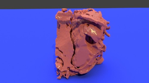
Left Kidney with Tumor - stl file processed
By kminars
Left Kidney with Tumor - stl file processed
23 downloads
(0 reviews)0 comments
Updated
-

Free
(0 reviews)0 comments
Updated
-

Free
(0 reviews)0 comments
Updated
-

Free
CT SCAN - CHEST-PELVIC - 10-27-16 - processed
By AABERNETHY
CT SCAN - CHEST-PELVIC - 10-27-16 - processed, ribs, .stl, 3d, model, printable, abdomen, rectum, pelvis, iliac, bone, ischium, pubis, gluteus, heart, .stl,
5 downloads
(0 reviews)0 comments
Updated


-
File Reviews
-
File Comments
-
Recent Forum Posts
-
 Hello everyone, I hope this message finds you well. I am reaching out to our community in hopes of finding assistance with a project I'm currently undertaking. Specifically, I am in need of a detailed 3D model of the temporomandibular joint (TMJ). If anyone in our community has access to or expertise in creating high-quality 3D models, particularly of the temporomandibular joint, I would greatly appreciate any assistance you can offer. Whether you have a model readily available or can provide guidance on where to find one, your contribution would be invaluable to my project. Thank you in advance for your assistance and support.
 -

-

-

-

By Georgecilia · Posted
With the outbreak of the coronavirus pandemic, many industries have been affected, including healthcare. One area where 3D printing has shown promise is in the production of medical supplies such as face shields, ventilator parts, and even swabs for testing. The ability to rapidly prototype and produce these items using 3D printing technology has allowed for a quicker response to the growing demand for essential medical equipment.
-


