
-
Welcome to embodi3D Downloads! This is the largest and fastest growing library of 3D printable anatomic models generated from real medical scans on the Internet. A unique scientific resource, most of the material is free. Registered members can download, upload, and sell models. To convert your own medical scans to a 3D model, take a look at democratiz3D, our free and automated conversion service.
Alert (6/17/22) - The democratiz3D scan-to-model conversion app is down due to a technical issue. We are working on a solution.
Congenital Heart Defects
A collection of files for 3D printing demonstrating congenital heart defects.
69 files
-

Free
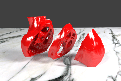
3D Printable Heart Model with Tetrology of Fallot
By embodi3d
This three-part 3D printed heart is from a CT scan of a 4-year-old infant with Tetrology of Fallot, a congentital heart defect and the most common cause of blue baby syndrome. It is characterized by stenosis (narrowing) of the pulmonary artery, an abnormal defect of the ventricular septum, an "overriding" aorta, and hypertrophy of the right ventricle. This patient has had a corrective surgery called Blalock-Taussig shunt.
The three STL files have been zipped and available for download. Alternatively, one STL file representing the whole model is also available for download. The three part model has holes for magnets, which can be used to connect and separate the pieces. The magnets can be found on a site which specializes in rare earth magnets.
The model is provided for distribution on Embodi3D with the permission of the author, pediatric cardiologist Dr. Matthew Bramlet, MD, and is part of the Heart Library. We thank Dr. Bramlet and all others who are working to help children with congenital heart problems lead normal and happy lives.
It is distributed by Dr. Bramlet under the Creative Commons license Attribution-NonCommercial-NoDerivs. Please respect the terms of the licensing agreement.
A US quarter is shown for scale in the images below.
467 downloads
- congenital heart disease
- Tetrology of Fallot
- (and 5 more)
(1 review)0 comments
Updated
-
Free
Atrial Septal Defect (ASD) and Mild Coarctation STL Files for 3D Printing
By embodi3d
There are four STL files for 3D printing demonstrating a moderate secundum atrial septal defect (ASD) and a mild coarctation. An atrial septal defect is a birth defect of the heart in which there is a hole in the wall (septum) that divides the upper chambers of the heart (atria). A hole can vary in size and may close on its own or may require surgery. If one of these openings does not close, a hole is left, and it is called an atrial septal defect. The hole increases the amount of blood that flows through the lungs and over time, it may cause damage to the blood vessels in the lungs. Damage to the blood vessels in the lungs may cause problems in adulthood, such as high blood pressure in the lungs and heart failure. Other problems may include abnormal heartbeat, and increased risk of stroke.
MRI obtained for evaluation of distal arch and pulmonary veins due to findings of pulmonary overcirculation out of proportion to typical ASD pathophysiology.
The MRI provided a complete anatomic overview and quantified the right sided enlargement from the 2:1 shunt through the ASD. Due to saturation band nulling of blood returning through the right sided pulmonary veins, there was excellent definition of the ASD due to the "dark" blood mixing with the "bright" blood and outlining the borders of the ASD which transfers to the model very well. Please keep in mind, that the model represents a heart in end-systole rather than diastole.
Disclaimer:
The available model has been validated to demonstrate the case’s pathologic features on a Z450 3D printer, (3DSystems, Circle Rock Hill, South Carolina)(or other printer as appropriate). While the mask applied to the original DICOM images accurately represents the anatomic features, some anatomic detail may be lost due to thin walled structures or inadequate supporting architecture; while other anatomic detail may be added due to similar limitations resulting in bleeding of modeling materials into small negative spaces. However, intracardiac structures, relationships, and pathologic features represent anatomic findings to scale and in high detail.
Credit:
The model is provided for distribution on Embodi3D with the permission of the author, pediatric cardiologist Dr. Matthew Bramlet, MD, and is part of the Congenital Heart Defects library. We thank Dr. Bramlet and all others who are working to help children with congenital heart problems lead normal and happy lives.
It is distributed by Dr. Bramlet under the Creative Commons license Attribution-NonCommercial-NoDerivs. Please respect the terms of the licensing agreement.
956 downloads
- secundum atrial septal defect
- atrial septal defect
- (and 5 more)
(1 review)0 comments
Submitted
-

$80
3D Model of Heart (2.3.4.5 chamber view) - 4 pack
By micamaca
Are you looking for a heart-stoppingly good way to impress your friends and colleagues? Look no further than our 3D models of heart apical 5 chamber view, perfect for anyone who loves anatomy, medicine, or just plain old cool stuff. With intricate details and lifelike accuracy, our models are the perfect addition to any desk or display. Impress your cardiologist friends or use it to educate your patients on the inner workings of their most vital organ. Plus, with the ability to print in a variety of materials, you can make your heart model as realistic or as colorful as you'd like. So what are you waiting for? Grab a 3D heart model today and show off your love for all things cardiac!
This pack includes:
3D Model of Heart (apical 2 chamber plane) 3D Model of Heart (apical 3 chamber plane) 3D Model of Heart (apical 4 chamber plane) 3D Model of Heart (apical 5 chamber plane)32 downloads
- heart
- cardiovascular
- (and 8 more)
(0 reviews)0 comments
Submitted
-

$99.99
Congenital Heart Disease - 7pack
By micamaca
Greetings! Have you ever wanted to explore the intricate world of congenital heart diseases? Our 3D model of a heart with congenital heart diseases is the perfect way to do just that. Congenital heart disease refers to any structural abnormality of the heart that is present at birth, and affects approximately 1 in every 100 babies born. These defects can range from simple to complex, and can cause a wide variety of symptoms. Our highly-detailed 3D model allows you to visualize and understand the complex anatomy of congenital heart diseases in a way that traditional textbooks simply cannot match. With the ability to 3D model and print the heart, you can hold a physical representation of the defect and study it in detail. This model is perfect for medical students, doctors, or anyone with an interest in cardiology. So why settle for a 2D image when you can hold a physical model of a congenital heart disease in your hands? Buy our 3D model today and gain a deeper understanding of these fascinating conditions.
With this Congenital Heart disease Pack you will get 7 different 3D models.
This Congenital Heart Disease Pack includes:
1. Atrial Septal Defect (ASD):
2. Ventricular Septal Defect (VSD)
3. Bicuspid Aortic Valve (BAV)
4. Common arterial trunk (CAT)
5. Partial Anomalous Pulmonary Venous Connection (PAPVC)
6. Tetralogy of Fallot (TOF)
7. Transposition of the Great Arteries (TOGA)
For each congenital heart disease following file formats are included: stl, obj, fbx
Short YT video:
11 downloads
- congenital
- congenital heart
- (and 21 more)
(0 reviews)0 comments
Updated
-

$25
3d model of heart with partial anomalous pulmonary venous connection
By micamaca
Looking for a detailed and accurate 3D model of partial anomalous pulmonary venous connection (PAPVC) to enhance your medical education, research, or patient communication? Look no further than our 3D model.
PAPVC is a rare congenital heart defect where one or more of the pulmonary veins drain into the right atrium, instead of the left atrium. This condition can lead to a range of symptoms and complications, including fatigue, shortness of breath, and an increased risk of heart failure. Our 3D model of PAPVC is designed to help you better understand the complex anatomy of this condition, and visualize its associated abnormalities.
Our 3D model is created using the latest in 3D printing technology, and is made from high-quality, durable materials to ensure longevity and ease of use. It accurately represents the intricate anatomy of PAPVC, including the location of the anomalous pulmonary veins, and the associated abnormalities such as atrial septal defects (ASD) and patent foramen ovale (PFO). It is an ideal tool for medical education and training, surgical planning, and patient communication.
Whether you are a medical professional, student, or patient, our 3D model of PAPVC is a valuable resource that can help you better understand this complex condition, and communicate its complexities to others. Don't settle for less than the best - order your 3D model today and take the first step towards improved medical education, research, and patient care.
Triangles: 210.2k
Vertices: 97.1k
Files included: obj, stl, fbx, dae
Short YT video:
0 downloads
(0 reviews)0 comments
Updated
-

$25
3d model of heart with common arterial trunk
By micamaca
Looking for an accurate and detailed 3D model of the common arterial trunk to enhance your medical education, research, or patient communication? Our 3D model is the perfect tool for you.
The common arterial trunk is a congenital heart defect that occurs when the aorta and pulmonary artery arise from a single trunk, rather than separately as in a normal heart. This condition can lead to a variety of serious complications, including heart failure, pulmonary hypertension, and cyanosis. Our 3D model of the common arterial trunk is designed to help you better understand the anatomy of this complex condition, and visualize its associated abnormalities.
Our 3D model is created using the latest in 3D printing technology, and is made from high-quality, durable materials to ensure longevity and ease of use. It accurately represents the intricate anatomy of the common arterial trunk, including the aortic and pulmonary valves, and the branching of the pulmonary artery and aorta. It is an ideal tool for medical education and training, surgical planning, and patient communication.
Whether you are a medical professional, student, or patient, our 3D model of the common arterial trunk is a valuable resource that can help you better understand this complex condition, and communicate its complexities to others. Don't settle for less than the best - order your 3D model today and take the first step towards improved medical education, research, and patient care.
Files included: obj, stl, dae
Triangles: 207.2k
Vertices: 103.1k
Short YT video:
0 downloads
(0 reviews)0 comments
Updated
-

$25
3d model of heart with bicuspid aortic valve
By micamaca
Are you looking for a unique and anatomically accurate 3D model to use in your medical education, research, or patient communication? Look no further than our detailed 3D model of the bicuspid aortic valve. The bicuspid aortic valve is a common congenital heart defect that affects up to 2% of the population, and is characterized by the presence of only two leaflets in the aortic valve instead of the usual three. This can lead to complications such as aortic stenosis, aortic regurgitation, and aneurysms, which may require surgical intervention. Our 3D model accurately represents the complex anatomy of the bicuspid aortic valve, including the leaflets, cusps, and sinuses of Valsalva, and can be used to visualize the condition and its associated abnormalities. Our 3D model is created using the latest in 3D printing technology, and is made from high-quality, durable materials to ensure longevity and ease of use. It can be used for a variety of purposes, including medical education and training, surgical planning, and patient communication. Whether you are a medical professional, student, or patient, our 3D model of the bicuspid aortic valve is an invaluable tool that can help you better understand this complex condition and its associated complications. So why wait? Order your 3D model today and take the first step towards improved medical education, research, and patient care.
Formats included: stl, obj, fbx, dae
Triangles: 197.2k
Vertices: 98.6k
Short YT video:
0 downloads
(0 reviews)0 comments
Updated
-

$25
3d model of the heart with tetralogy of Fallot parasternal long axis
By micamaca
Greetings! Are you interested in learning about congenital heart defects? Our 3D model of a heart with tetralogy of fallot (TOF) is the perfect way to explore this fascinating condition. Our highly-detailed 3D model allows you to visualize and understand the complex anatomy of an TOF in a way that traditional textbooks simply cannot match. With the ability to 3D print the model, you can hold a physical representation of the defect and study it in detail. This model is perfect for medical students, doctors, or anyone with an interest in cardiology. So why settle for a 2D image when you can hold a physical model of an atrial septal defect in your hands? Buy our 3D model today and gain a deeper understanding of this fascinating condition.
Tetralogy of Fallot is a congenital heart defect that occurs during fetal development, affecting the heart's structure and function. It is a combination of four defects that occur together: a ventricular septal defect (a hole between the two lower chambers of the heart), pulmonary stenosis (narrowing of the pulmonary valve and artery), overriding aorta (the aorta is positioned over both the left and right ventricles), and right ventricular hypertrophy (thickening of the right ventricle wall due to increased workload). These defects result in reduced blood flow to the lungs and inadequate oxygen supply to the body.
Formats: dae, obj, stl
Triangles: 196,500
Vertices: 98,300
Short YT video:
33 downloads
- tetralogy of fallot
- heart
- (and 17 more)
(0 reviews)0 comments
Submitted
-

$20
3d model of pulmonary arteries (Fontan Procedure)
By micamaca
Single ventricle anomalies are rare congenital heart defects in which one of the ventricles (or chambers) of the heart does not develop properly.
The Fontan procedure is the last in a series of operations that tries to establish a near-normal circulation of blood for patients with functional single-ventricle physiology.
The goal of the procedure is to separate systemic and pulmonary blood flow by directing systemic venous return through the Fontan connection to the pulmonary arteries and the lungs without ventricular contribution.
Following the procedure, pulmonary blood flow is completely passive and dependent on pressure gradients, resulting in complex postoperative cardiopulmonary interactions.
The model is provided in .stl, and .obj file formats.
Additional supports are needed for printing.
Initial size: 189.8 x 127.3 x 155.2 mm
Estimated printing time with support is around 30 h and 38 minutes (with 0.15 mm precision).
3 downloads
- cardiovascular
- cardiovascular system
- (and 11 more)
(0 reviews)0 comments
Submitted
-

$20
3d model of pulmonary arteries (Fontan Procedure)
By micamaca
Single ventricle anomalies are rare congenital heart defects in which one of the ventricles (or chambers) of the heart does not develop properly.
The Fontan procedure is the last in a series of operations that tries to establish a near-normal circulation of blood for patients with functional single-ventricle physiology.
The goal of the procedure is to separate systemic and pulmonary blood flow by directing systemic venous return through the Fontan connection to the pulmonary arteries and the lungs without ventricular contribution.
Following the procedure, pulmonary blood flow is completely passive and dependent on pressure gradients, resulting in complex postoperative cardiopulmonary interactions.
The model is provided in .stl, and .obj file formats.
Additional supports are needed for printing.
Initial size: 159.0 x 85.1 x 121.2 mm
Estimated printing time with support is around 17 h (with 0.15 mm precision).
2 downloads
- cardiovascular
- cardiovascular system
- (and 11 more)
(0 reviews)0 comments
Submitted
-

$20
3d model of pulmonary arteries (Fontan Procedure)
By micamaca
Single ventricle anomalies are rare congenital heart defects in which one of the ventricles (or chambers) of the heart does not develop properly.
The Fontan procedure is the last in a series of operations that tries to establish a near-normal circulation of blood for patients with functional single-ventricle physiology.
The goal of the procedure is to separate systemic and pulmonary blood flow by directing systemic venous return through the Fontan connection to the pulmonary arteries and the lungs without ventricular contribution.
Following the procedure, pulmonary blood flow is completely passive and dependent on pressure gradients, resulting in complex postoperative cardiopulmonary interactions.
The model is provided in .stl, and .obj file formats.
Additional supports are needed for printing.
Initial size: 151.1 x 83.1 x 112.1 mm
Estimated printing time with support is around 14 h and 58 minutes (with 0.15 mm precision).
2 downloads
- cardiovascular
- cardiovascular system
- (and 10 more)
(0 reviews)0 comments
Submitted
-

$20
3d model of pulmonary arteries (Fontan Procedure)
By micamaca
Single ventricle anomalies are rare congenital heart defects in which one of the ventricles (or chambers) of the heart does not develop properly.
The Fontan procedure is the last in a series of operations that tries to establish a near-normal circulation of blood for patients with functional single-ventricle physiology.
The goal of the procedure is to separate systemic and pulmonary blood flow by directing systemic venous return through the Fontan connection to the pulmonary arteries and the lungs without ventricular contribution.
Following the procedure, pulmonary blood flow is completely passive and dependent on pressure gradients, resulting in complex postoperative cardiopulmonary interactions.
The model is provided in .stl, and .obj file formats.
Additional supports are needed for printing.
Initial size: 131.1 x 102.7 x 106.0 mm
Estimated printing time with support is around 15 h and 31 minutes (with 0.15 mm precision).
2 downloads
- cardiovascular
- cardiovascular system
- (and 11 more)
(0 reviews)0 comments
Submitted
-

$20
3d model of pulmonary arteries (Fontan Procedure)
By micamaca
Single ventricle anomalies are rare congenital heart defects in which one of the ventricles (or chambers) of the heart does not develop properly.
The Fontan procedure is the last in a series of operations that tries to establish a near-normal circulation of blood for patients with functional single-ventricle physiology.
The goal of the procedure is to separate systemic and pulmonary blood flow by directing systemic venous return through the Fontan connection to the pulmonary arteries and the lungs without ventricular contribution.
Following the procedure, pulmonary blood flow is completely passive and dependent on pressure gradients, resulting in complex postoperative cardiopulmonary interactions.
The model is provided in .stl, and .obj file formats.
Additional supports are needed for printing.
2 downloads
- cardiovascular
- cardiovascular system
- (and 12 more)
(0 reviews)0 comments
Submitted
-

$20
3d model of pulmonary arteries (Fontan Procedure)
By micamaca
Single ventricle anomalies are rare congenital heart defects in which one of the ventricles (or chambers) of the heart does not develop properly.
The Fontan procedure is the last in a series of operations that tries to establish a near-normal circulation of blood for patients with functional single-ventricle physiology.
The goal of the procedure is to separate systemic and pulmonary blood flow by directing systemic venous return through the Fontan connection to the pulmonary arteries and the lungs without ventricular contribution.
Following the procedure, pulmonary blood flow is completely passive and dependent on pressure gradients, resulting in complex postoperative cardiopulmonary interactions.
The model is provided in .stl, and .obj file formats.
Additional supports are needed for printing.
2 downloads
- cardiovascular
- cardiovascular system
- (and 13 more)
(0 reviews)0 comments
Submitted
-

$20
3d model of pulmonary arteries (Fontan Procedure)
By micamaca
Single ventricle anomalies are rare congenital heart defects in which one of the ventricles (or chambers) of the heart does not develop properly.
The Fontan procedure is the last in a series of operations that tries to establish a near-normal circulation of blood for patients with functional single-ventricle physiology.
The goal of the procedure is to separate systemic and pulmonary blood flow by directing systemic venous return through the Fontan connection to the pulmonary arteries and the lungs without ventricular contribution.
Following the procedure, pulmonary blood flow is completely passive and dependent on pressure gradients, resulting in complex postoperative cardiopulmonary interactions.
The model is provided in .stl, and .obj file formats.
Additional supports are needed for printing.
2 downloads
(0 reviews)0 comments
Updated
-

$20
3d model of pulmonary arteries (Fontan Procedure)
By micamaca
Single ventricle anomalies are rare congenital heart defects in which one of the ventricles (or chambers) of the heart does not develop properly.
The Fontan procedure is the last in a series of operations that tries to establish a near-normal circulation of blood for patients with functional single-ventricle physiology.
The goal of the procedure is to separate systemic and pulmonary blood flow by directing systemic venous return through the Fontan connection to the pulmonary arteries and the lungs without ventricular contribution.
Following the procedure, pulmonary blood flow is completely passive and dependent on pressure gradients, resulting in complex postoperative cardiopulmonary interactions.
The model is provided in .stl, and .obj file formats.
Additional supports are needed for printing.
Initial size: 170.8 x 149.7 x 155.7 mm
Estimated printing time with support is around 27 h and 32 minutes (with 0.15 mm precision).
2 downloads
- cardiovascular
- cardiovascular system
- (and 13 more)
(0 reviews)0 comments
Submitted
-

$20
3d model of pulmonary arteries (Fontan Procedure)
By micamaca
Single ventricle anomalies are rare congenital heart defects in which one of the ventricles (or chambers) of the heart does not develop properly.
The Fontan procedure is the last in a series of operations that tries to establish a near-normal circulation of blood for patients with functional single-ventricle physiology.
The goal of the procedure is to separate systemic and pulmonary blood flow by directing systemic venous return through the Fontan connection to the pulmonary arteries and the lungs without ventricular contribution.
Following the procedure, pulmonary blood flow is completely passive and dependent on pressure gradients, resulting in complex postoperative cardiopulmonary interactions.
The model is provided in stl, and obj file formats.
Additional supports are needed to print the various components.
2 downloads
- cardiovascular
- cardiovascular system
- (and 17 more)
(0 reviews)0 comments
Updated
-

$20
3d model of pulmonary arteries (Fontan Procedure)
By micamaca
Single ventricle anomalies are rare congenital heart defects in which one of the ventricles (or chambers) of the heart does not develop properly.
The Fontan procedure is the last in a series of operations that tries to establish a near-normal circulation of blood for patients with functional single-ventricle physiology.
The goal of the procedure is to separate systemic and pulmonary blood flow by directing systemic venous return through the Fontan connection to the pulmonary arteries and the lungs without ventricular contribution.
Following the procedure, pulmonary blood flow is completely passive and dependent on pressure gradients, resulting in complex postoperative cardiopulmonary interactions.
The model is provided in stl, and obj file formats.
Additional supports are needed to print the various components.
2 downloads
- cardiovascular
- cardiovascular system
- (and 14 more)
(0 reviews)0 comments
Updated
-

Free
Coarctation of aorta - stl file processed
By Lilian
Coarctation of aorta - stl file processed
Have embodi3D 3D print this model for you. This file was created with democratiz3D. Automatically create 3D printable models from CT scans.6 downloads
(0 reviews)0 comments
Submitted
-

$25
Model of human heart with hypertrophic cardiomyopathy generated from real patient
By micamaca
This 3D printable model of a human heart with hypertrophic cardiomyopathy was generated from an real life patient CT scan.
It includes 7 parts including the aorta, left and right atrium, left and right ventricles, and pulmonary artery.
The model demonstrates the detailed anatomy of the various blood pools of the human heart.
The model comprise of 9 .stl files: whole heart and 7 separate heart structures
Mesh integrity: manifold STL (watertight)
Additional supports are needed to print the various components.
15 downloads
- cardiovascular
- cardiovascular system
- (and 6 more)
(0 reviews)0 comments
Updated
-

$25
Model of human heart with mirror dextrocentric generated from real patient
By micamaca
This 3D printable model of a human heart with mirror dextrocentric was generated from an real life patient CT scan.
It includes 8 parts including the aorta, left and right atrium, left and right ventricles, and pulmonary artery.
The model demonstrates the detailed anatomy of the various blood pools of the human heart.
The model comprise of 9 .stl files: whole heart and 8 separate heart structures
Mesh integrity: manifold STL (watertight)
Additional supports are needed to print the various components.
8 downloads
- mirror dextrocentric+
- cardiovascular
- (and 8 more)
(0 reviews)0 comments
Updated
-

$25
Model of human heart with anomalous pulmonary venous drainage (APVC) generated from real patient
By micamaca
This 3D printable model of a human heart with anomalous pulmonary venous drainage (APVC) was generated from an real life patient CT scan by clinical expert.
It includes 7 parts including the aorta, left and right atrium, left and right ventricles, and pulmonary artery.
The model demonstrates the detailed anatomy of the various blood pools of the human heart.
The model comprise of 8 .stl files: whole heart and 7 separate heart structures
Mesh integrity: manifold STL (watertight)
Additional supports are needed to print the various components.
7 downloads
- cardiovascular system
- cardiovascular
- (and 6 more)
(0 reviews)0 comments
Updated
-

$25
Model of human heart with atrial septal defect (ASD) generated from real patient
By micamaca
Heart affected by ASD has unexpected connection between left and right atrium.
This 3D printable model of a human heart with atrial septal defect was generated from an real life patient CT scan.
It includes 7 parts including the aorta, left and right atrium, left and right ventricles, and pulmonary artery.
The model demonstrates the detailed anatomy of the various blood pools of the human heart.
The model comprise of 8 .stl files: whole heart and 7 separate heart structures
Mesh integrity: manifold STL (watertight)
Additional supports are needed to print the various components.
15 downloads
- cardiovascular system
- cardiac
- (and 6 more)
(0 reviews)0 comments
Updated
-

$25
Model of human heart with atrio-ventricular septal defect (AVSD) generated from real patient
By micamaca
Atrio-ventricular septal defect (AVSD) is a combination of ASD and VSD, and the it has unexpected connections between left atrium/left ventricle and right atrium/right ventricle.
This 3D printable model of a human heart with atrio-ventricular septal defect was generated from an real life patient CT scan by clinical expert.
It includes 7 parts including the aorta, left and right atrium, left and right ventricles, and pulmonary artery.
The model demonstrates the detailed anatomy of the various blood pools of the human heart.
The model comprise of 8 .stl files: whole heart and 7 separate heart structures
Mesh integrity: manifold STL (watertight)
Additional supports are needed to print the various components.
15 downloads
- cardiovascular system
- cardiova
- (and 6 more)
(0 reviews)0 comments
Updated
-

$25
Model of human heart with co-arctation (CA) generated from real patient
By micamaca
This 3D printable model of a human heart with co-arctation (CA) was generated from an real life patient CT scan by clinical expert.
It includes 7 parts including the aorta, left and right atrium, left and right ventricles, and pulmonary artery.
The model demonstrates the detailed anatomy of the various blood pools of the human heart.
The model comprise of 8 .stl files: whole heart and 7 separate heart structures
Mesh integrity: manifold STL (watertight)
Additional supports are needed to print the various components.
7 downloads
- cardiovascular
- cardiovascular system
- (and 6 more)
(0 reviews)0 comments
Updated


-
File Reviews
-
File Comments
-
Recent Forum Posts
-
 Hello everyone, I hope this message finds you well. I am reaching out to our community in hopes of finding assistance with a project I'm currently undertaking. Specifically, I am in need of a detailed 3D model of the temporomandibular joint (TMJ). If anyone in our community has access to or expertise in creating high-quality 3D models, particularly of the temporomandibular joint, I would greatly appreciate any assistance you can offer. Whether you have a model readily available or can provide guidance on where to find one, your contribution would be invaluable to my project. Thank you in advance for your assistance and support.
 -

-

-

-

By Georgecilia · Posted
With the outbreak of the coronavirus pandemic, many industries have been affected, including healthcare. One area where 3D printing has shown promise is in the production of medical supplies such as face shields, ventilator parts, and even swabs for testing. The ability to rapidly prototype and produce these items using 3D printing technology has allowed for a quicker response to the growing demand for essential medical equipment.
-


