
Invasive Heart Surgery Avoided as Advances in 3D Printing Improve Safety of Transcatheter Aortic Valve Replacement (TAVR)

Entry posted by Dr. Mike ·
4,370 views

3D printing is revolutionizing the treatment of aortic stenosis, as reported by researchers from St. Joseph's Hospital in Phoenix, Arizona and presented at the 2014 Radiological Society of North American (RSNA) meeting. Aortic stenosis is a deadly condition where the valve that connects the heart to the aorta does not open properly. The aortic valve, as it is called, is designed to open freely to allow blood pumped from the heart to move in a forward direction into the aorta, the main artery of the body. At the end of a heart contraction, the valve closes to prevent the blood from flowing backwards. In patients with aortic stenosis, the valve fails to open during contraction, thus preventing blood from flowing forward during cardiac contraction (systole). The heart compensates by squeezing harder and harder to maintain adequate forward flow. Eventually, the heart becomes too strained and a variety of severe complications can ensure, including heart attack, heart failure, syncope (fainting), and sudden death. In patients with severe aortic stenosis there is a 50% chance of dying within two years if untreated.
Traditional treatment of aortic stenosis involved open heart surgery to replace the valve. This is a major operation and involves sawing through the breast bone to open the chest and gain access to the heart. As one can imagine, the procedure is risky and recovery takes a long time. Many patients who are too ill or weak and are not eligible for the surgery.
Fortunately, there is a new procedure where a prosthetic aortic valve can be implanted by navigating a plastic tube, or catheter, to the heart through a puncture in the artery of the hip or sometimes via a direct puncture through the apex of the heart. This procedure, called a Transcatheter Aortic Valve Replacement, or TAVR, promises to make aortic valve replacement minimally invasive, that is doable through a small hole instead of requiring open surgery. Recovery time is significantly less and patients who are ineligible for conventional surgery can often get TAVR.
The major problem with TAVR is that it can be very difficult to accurately place the prosthetic valve through the catheter, and poor placement can result in devastating consequences. Unlike open surgery, where the surgeon has direct access to the diseased heart valve, with TAVR the physician must work through the catheter. The valve is loaded on a collapsed metal mesh called a stent. The stent is then inched forward through the catheter into the proper position. The position is checked with x-ray and then the stent is expanded -- hopefully in the correct position relative to the diseased aortic valve, pushing it aside and replacing it with the new prosthetic valve. Once the prosthetic valve is expanded it cannot be retrieved or repositioned, and millimeters matter. Valves come in different sizes. If the valve is the wrong size or malpositioned by even a few millimeters it can cover the coronary arteries and lead to heart attack, cause blood clots to form leading to stroke, or even come free and cause tearing of the aorta.
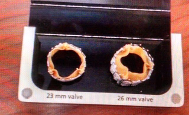
Different sized TAVR aortic valves
This is where 3D printing comes in. The Arizona researchers used contrast enhanced computed tomography (CT) scans to gather precise data on the anatomical structure of the heart, aorta, and aortic valve from three patients with severe aortic stenosis, and manufactured precise 3D printed replicas of the aortas. Then they tested various valve sizes and position to see what had the best anatomic fit. They then did CT scans of the prosthetic valves within the 3D models and compared those to postoperative CT scans of the actual patients after TAVR, and found that the pre-surgical test fitting in the 3D printed models accurately predicted how the valves would perform in real patients. Use of the 3D printed models helped the surgeons choose the correct size valve and positioning prior to the surgery, thus reducing the risk of he TAVR procedure.
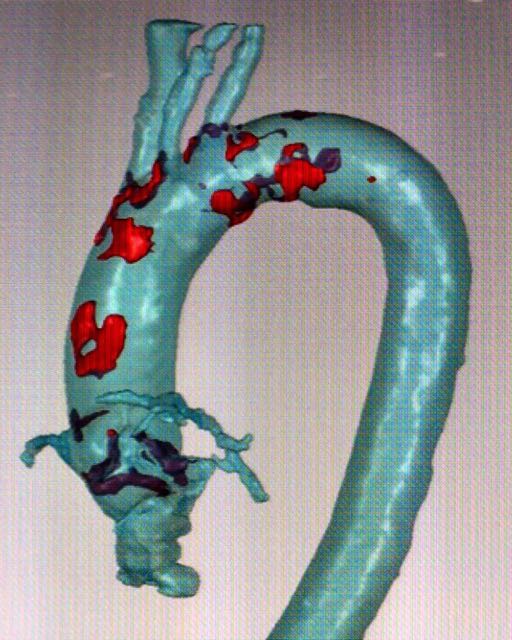
Digital rendering of the aorta
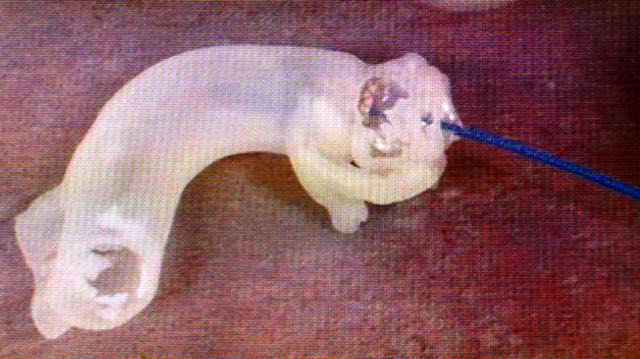
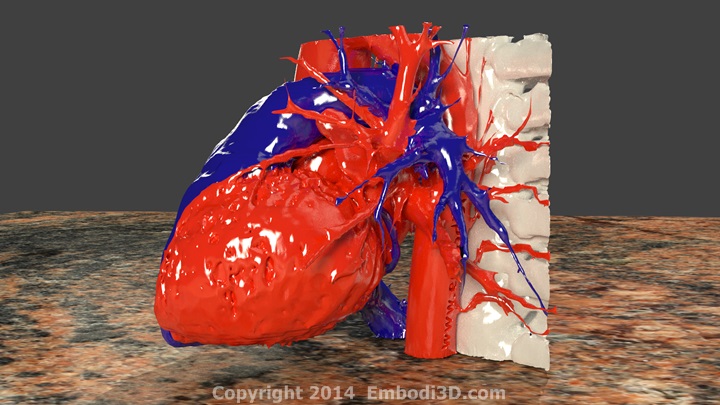
Testing the implantable valve in the 3D printed model by deploying it through a catheter (blue)
I have personally used customized 3D printed models I designed to test wires and catheters prior to complex mesenteric artery aneurysm treatments (publication forthcoming), and I can tell you that knowing your wires and catheters will work before the procedure is far better than figuring it out with trial and error during the procedure. Presurgical testing with 3D printed models is here to stay.
If you are interested in learning more about applications of 3D printing in medicine or how to make your own 3D models, please register as a member (it's free and only takes a minute!) and join the community of medical professionals who are all trying to build the future of 3D printing in medicine. Ask a question, start a discussion, or download free 3D printable models to make on your own. I will put some links to a few of the free downloadable models related to this article below.
FREE 3D PRINTABLE DOWNLOADS
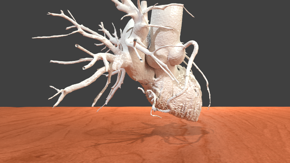
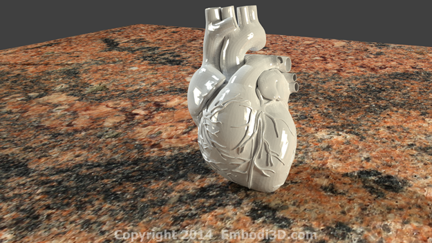
Heart and pulmonary artery tree
http://www.embodi3d.com/files/file/59-heart-and-pulmonary-artery-tree-from-ct-angiogram/
Human heart
http://www.embodi3d.com/files/file/27-human-heart/
Human heart #2
http://www.embodi3d.com/files/file/35-3d-printable-human-heart/


0 Comments
Recommended Comments
There are no comments to display.
Create an account or sign in to comment
You need to be a member in order to leave a comment
Create an account
Sign up for a new account in our community. It's easy!
Register a new accountSign in
Already have an account? Sign in here.
Sign In Now